Search anything and hit enter
  • Teams
  • Members
  • Projects
  • Events
  • Calls
  • Jobs
  • publications
  • Software
  • Tools
  • Network
  • Equipment

A little guide for advanced search:

  • Tip 1. You can use quotes "" to search for an exact expression.
    Example: "cell division"
  • Tip 2. You can use + symbol to restrict results containing all words.
    Example: +cell +stem
  • Tip 3. You can use + and - symbols to force inclusion or exclusion of specific words.
    Example: +cell -stem
e.g. searching for members in projects tagged cancer
Search for
Count
IN
OUT
Content 1
  • member
  • team
  • department
  • center
  • program_project
  • nrc
  • whocc
  • project
  • software
  • tool
  • patent
  • Administrative Staff
  • Assistant Professor
  • Associate Professor
  • Clinical Research Assistant
  • Clinician Researcher
  • Department Manager
  • Full Professor
  • Graduate Student
  • Honorary Professor
  • Lab assistant
  • Non-permanent Researcher
  • Permanent Researcher
  • Pharmacist
  • PhD Student
  • Physician
  • Post-doc
  • Prize
  • Project Manager
  • Research Associate
  • Research Engineer
  • Retired scientist
  • Technician
  • Undergraduate Student
  • Veterinary
  • Visiting Scientist
  • Deputy Director of Center
  • Deputy Director of Department
  • Deputy Director of National Reference Center
  • Deputy Head of Facility
  • Director of Center
  • Director of Department
  • Director of Institute
  • Director of National Reference Center
  • Group Leader
  • Head of Facility
  • Head of Operations
  • Head of Structure
  • Honorary President of the Departement
  • Labex Coordinator
Content 2
  • member
  • team
  • department
  • center
  • program_project
  • nrc
  • whocc
  • project
  • software
  • tool
  • patent
  • Administrative Staff
  • Assistant Professor
  • Associate Professor
  • Clinical Research Assistant
  • Clinician Researcher
  • Department Manager
  • Full Professor
  • Graduate Student
  • Honorary Professor
  • Lab assistant
  • Non-permanent Researcher
  • Permanent Researcher
  • Pharmacist
  • PhD Student
  • Physician
  • Post-doc
  • Prize
  • Project Manager
  • Research Associate
  • Research Engineer
  • Retired scientist
  • Technician
  • Undergraduate Student
  • Veterinary
  • Visiting Scientist
  • Deputy Director of Center
  • Deputy Director of Department
  • Deputy Director of National Reference Center
  • Deputy Head of Facility
  • Director of Center
  • Director of Department
  • Director of Institute
  • Director of National Reference Center
  • Group Leader
  • Head of Facility
  • Head of Operations
  • Head of Structure
  • Honorary President of the Departement
  • Labex Coordinator
Search
Go back
Scroll to top
Share
Scientific Fields
Diseases
Organisms
Applications
Technique

About

The Image Analysis Hub is an open access, equal access core facility committed to offering bioimage analysis services to the scientific community.

What we do.

The IAH works mainly by collaborating scientifically with its users, trying to achieve a significant added-value to their Research.

Our services are deployed in four activities:

Infrastructure for autonomous image analysis.

We maintain and offer access to workstations dedicated to and tailored for image analysis software. This includes academic open-source software such as Fiji, Icy, ilastik, QuPath, … and commercial software like Imaris, Huygens and Arivis. They are under maintenance contract and regularly updated.

The workstations are located in a common analysis room in the François Jacob building (aquarium, room 26-01-17) and can be booked via our PPMS instance. We also offer access to virtual machines, that can be connected to remotely, on and off campus.

Walk-in support and trainings for questions involving image analysis.

Our users can consult us for bioimage analysis questions related to their project. We can also contribute to experimental design, with our colleagues from the imaging core facilities when required. We offer one-one training sessions on image analysis software, to address a specific question in a project. Finally, we teach general bioimage analysis techniques at introductory and advanced levels during yearly events such as bioimage analysis courses (like this one) and workshops or the PhD program.

Walk-in support happens mainly via our open-desk sessions. Every two weeks, on Thursday morning from 9:30am to 12:30am, the whole IAH and colleagues are gathered in the aquarium of the François Jacob building (room 26-01-17). We welcome users on a first-come first-served basis, and try to address their question immediately. Several analysts can work with one user, and we often invite research engineers from the campus to participate.

The open-desks sessions are the single entry point for all our services. If you want to request a collaborative project, a training or discuss what we could do to support your project, the one place to do so is the IAH open-desks. All the open-desk sessions for this year are listed on this page.

Build and deploy custom analysis tools for projects requiring special developments.

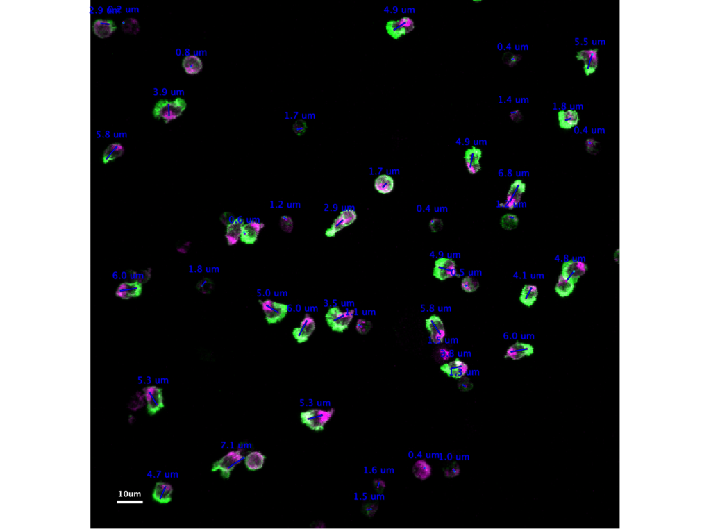
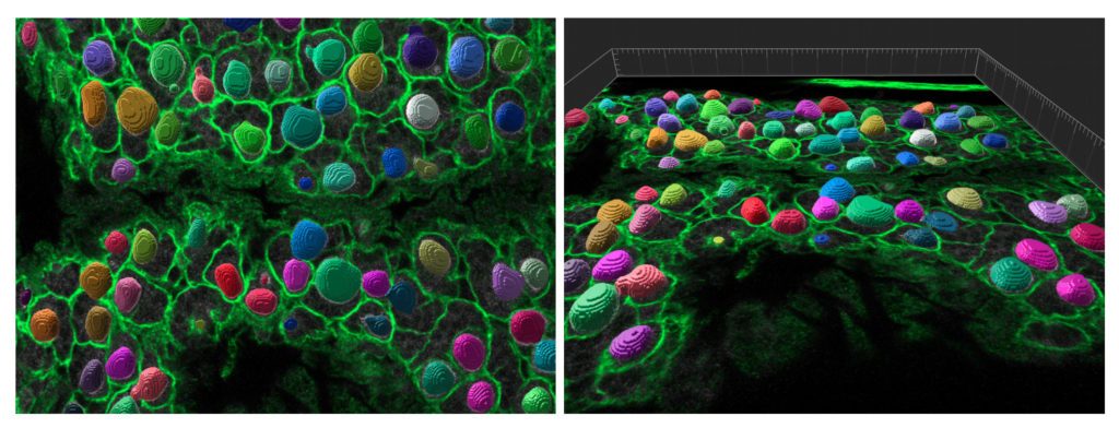
Our central activity consists in developing highly customized analysis pipelines or scientific tools tailored to address specifically a user project. This activity is important when a user project requires analysis that is not provided by the existing image analysis tools, or when they require modifications or integration in larger pipelines. The delivery of these services can take two forms:

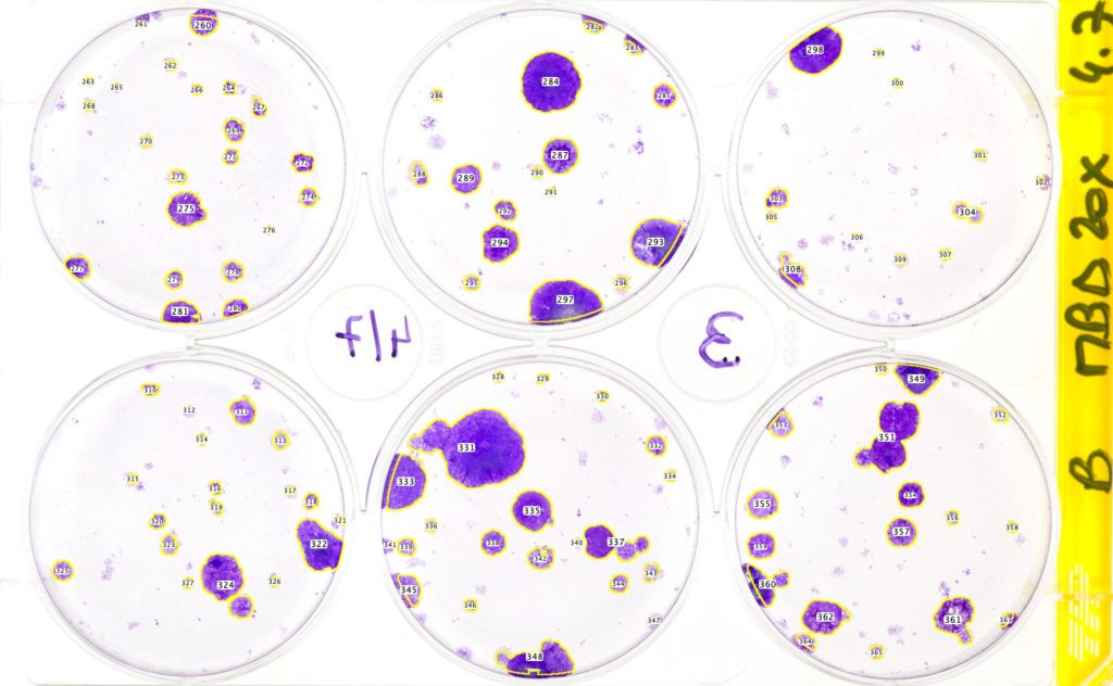
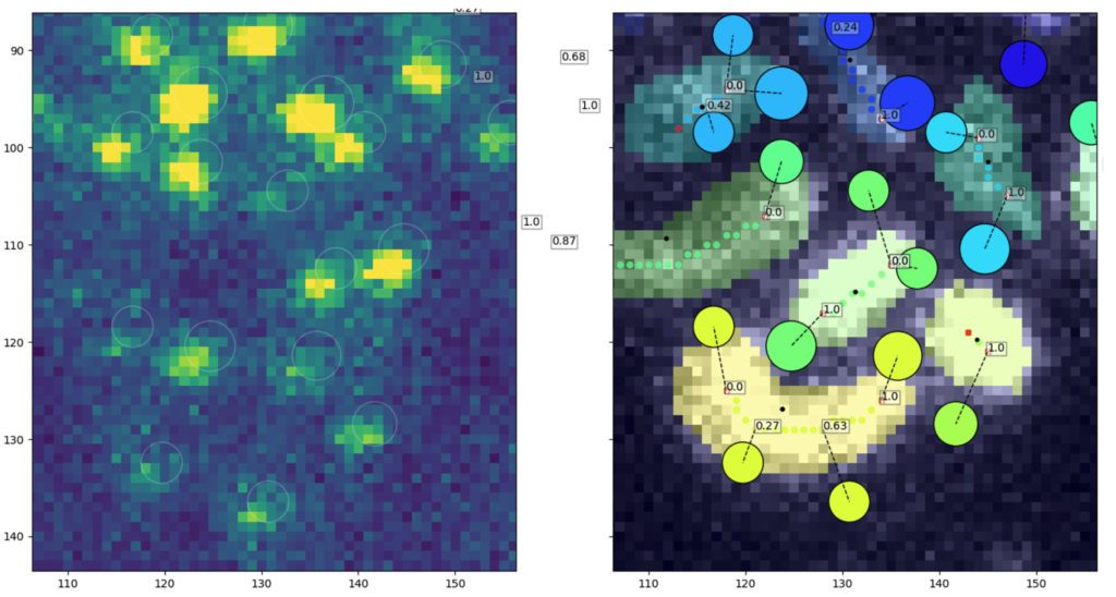
  • Quantification projects. A user would delegate an analysis task to us, a sub-part of their research project. Our goal then is produce scientific results from their images, up to the production of a figure for a publication, with the redaction of the associated Materials and Methods section.
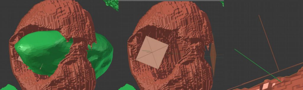
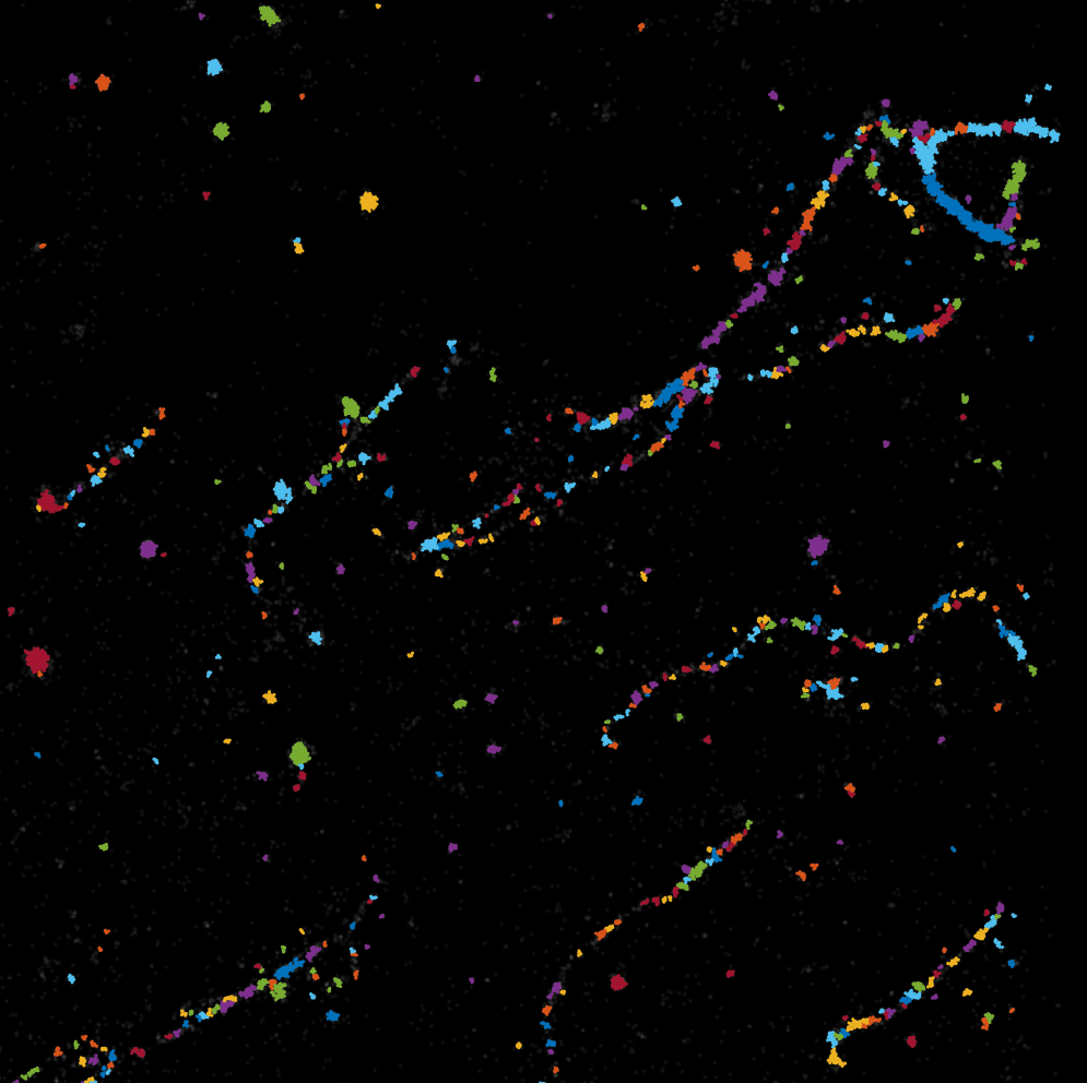
  • Custom development of analysis tools. We strive not to limit ourselves to be users of bioimage analysis software, but to be able to modify them and build new tools, tailored for the user project. Once built and validated, we deliver them to the user’s lab and teach their uses limitations.

Develop  software platforms for image analysis.

We are the developers of several image analysis platforms. For instance:

and we are involved in the core development of several software packages for bioimage analysis:

The scope of these software tools goes beyond a single user-project. They are developed and elaborated when used on multiple user projects and during dedicated engineer time. They are made to be extensible and modular, and are used later to quickly develop bespoke analysis pipeline for future user projects.

Obtained labels.

The IAH operates within a Quality Management System following the ISO-9001:2015 norm, and we achieved certification by the AFNOR in 2019, within the C2RT ISO-9001 certification. Following the ISO9001 certification, the IAH obtained the IBISA label in October 2019.

Logo AFNOR ISO9001


Equipments

Booking site

IAH workstation ‘Rammstein’ • Corsair • One

This PC serves as a platform for testing the software DIVA (Institut Pasteur, Institut Curie) that allows visualization of volumetric data (and not only) by means of Virtual Reality. Also, it is equipped with […]

IAH workstation ‘Tool’ • Intel-Nvidia-MSI • PC

This PC is meant for Image Analysis; it is equipped with a strong graphic processor, which allows usage of hardware-accelerated analysis software. CPU Intel 3.60 Ghz 6 Cores/12 Threads RAM 196 Gb DD4 GPU […]

Software

Courses

NEUBIAS Pasteur course on Bioimage Analysis

Paris, Institut Pasteur, from Tuesday the 30th of May to Friday June the 2nd. In-person only. Training school focused on Bioimage Analysis. Two parallel tracks: Early career investigators track: Learn to master the tools […]

2023-05-30 10:00:00 2023-06-02 00:00:00 Europe/Paris NEUBIAS Pasteur course on Bioimage Analysis Paris, Institut Pasteur, from Tuesday the 30th of May to Friday June the 2nd. In-person only. Training school focused on Bioimage Analysis. Two parallel tracks: Early career investigators track: Learn to master the tools […] 28 Rue du Docteur Roux, Paris, France

Microscopy – Principles and Applications of Fluorescence Microscopy

Fluorescence microscopy has revolutionized researchers’ ability to monitor cellular structures and dynamics. Over the last decades, the spectrum of applications of fluorescence microscopy has been constantly broadened by novel combinations of optics principles supported […]

2023-03-06 08:00:00 2023-03-17 19:00:00 Europe/Paris Microscopy – Principles and Applications of Fluorescence Microscopy Fluorescence microscopy has revolutionized researchers’ ability to monitor cellular structures and dynamics. Over the last decades, the spectrum of applications of fluorescence microscopy has been constantly broadened by novel combinations of optics principles supported […] 25-28 Rue du Dr Roux, 75015 Paris, France

Fundings

Partners

Publications

Download

Networks

NEUBIAS logo
NEUBIAS

Steering committee

The steering committee of the IAH includes three members, two from the Institut Pasteur, one external:

  • Perrine Paul-Gilloteaux. Scientific director of the MicroPICell facility, University of Nantes.
  • Romain Levayer. Head of the ‘Cell Death and Epithelial Homeostasis’ unit, Institut Pasteur, Paris.
  • Philippe Bastin. Head of the ‘Trypanosome Cell Biology’ unit, Institut Pasteur, Paris.

How to access the facility?

IAH open-desks.

First and foremost, come to our open-desks! Open-desk sessions aim at addressing the questions of the campus about image analysis. During 3 hours, once every 2 weeks, the IAH and a number of people from different labs and structures will be present to deal with your images, or general questions about image analysis. We also use them as the main entry point to the facility. Whether you want to start a project, request a training or just discuss an experiment, come to our open-desks Their planning and goals can be found on the IAH open-desk page: https://research.pasteur.fr/en/news/image-analysis-opendesk/

Emails and PPMS.

You can

  • make a project request;
  • make a training request

on our PPMS instance. Our activities are billed. Contact us for more information. We can be reached at iah at pasteur dot fr. If you are unhappy with our services or want to bring a point to our attention privately, you can do so iah.feedback at pasteur dot fr. Only the facility head and the administrative assistant receive these emails and they are treated confidentially.

Team chat.

If you like instant messaging, we have a public channel on the MS Teams instance of Pasteur. We use it to:

  • hold the remote open-desk sessions;
  • announce new tools we deploy;
  • or trainings we organize;
  • post interesting conference and external training announcements;
  • post papers we find interesting and comment them;
  • answer directly quick questions you post there.

Join the IAH channel on MS Teams.

Access policy.

We are part of the C2RT (Center for Technological Resources and Research), a center gathering the Technology and Service units (UTechS) and the technological core facilities of the campus. As such we follow the common guidelines and good practices. On top of this guidelines, our services require users to comply to the following policy:

1. Acknowledgements.

Users will acknowledge the IAH facility and its funding in any publication or communication, when its services (open-desks, advices, trainings, use of workstations, projects, …) have been used for the presented work. Please also let us know when you do so. Example acknowledgement sentence: “We acknowledge the help of Image Analysis Hub of the Institut Pasteur for this work.

2. Facility usage fees.

Users agree to pay the service fees related to the facility activities. Activities are billed based on hourly rate for academic users. Contact us for pricing.

3. Responsibility.

Users remain responsible for the storage and the integrity of the original image data they provide, and for the analysis results the IAH provides to them once they are delivered.

Pictures & Media

Measuring the polarization of T cells. Image M. Mastrogiovanni (Lymphocyte Cell Biology Unit, Institut Pasteur), J.-Y. Tinevez (IAH)
Identification and segmentation of stem cells in the drosophila post-embryonic brain. Image P. Speder (Brain Plasticity in Response to the Environment unit, Institut Pasteur), S. Rigaud (IAH).
Cell migration assay. Image G. Jacquemet (Cell Migration lab, Åbi Akademi University), J.-Y. Tinevez (IAH).
Colony counting on non-microscopy images. N. Gupta (Membrane Traffic and Cell Division unit), J.-Y. Tinevez (IAH).
Modeling a bacteria crossing a mitocondrium in electron microscopy images. Image Z. Uzum (Evolutionary Biology if the Microbial Cell, Institut Pasteur), D. Ershov (IAH).
Measuring the association of degradosome spots in Helicobacter pylori. Tejada-Arranz et al., 2020.
Automated doublet length measurement in super-resolution images. C. Leduc (Cell Polarity, Migration and Cancer unit), J.-Y. Tinevez (IAH)

Contact

Email: iah at pasteur dot fr Address: 25-28 Rue du Docteur Roux 75015, Paris France