Search anything and hit enter
  • Teams
  • Members
  • Projects
  • Events
  • Calls
  • Jobs
  • publications
  • Software
  • Tools
  • Network
  • Equipment

A little guide for advanced search:

  • Tip 1. You can use quotes "" to search for an exact expression.
    Example: "cell division"
  • Tip 2. You can use + symbol to restrict results containing all words.
    Example: +cell +stem
  • Tip 3. You can use + and - symbols to force inclusion or exclusion of specific words.
    Example: +cell -stem
e.g. searching for members in projects tagged cancer
Search for
Count
IN
OUT
Content 1
  • member
  • team
  • department
  • center
  • program_project
  • nrc
  • whocc
  • project
  • software
  • tool
  • patent
  • Administrative Staff
  • Assistant Professor
  • Associate Professor
  • Clinical Research Assistant
  • Clinical Research Nurse
  • Clinician Researcher
  • Department Manager
  • Dual-education Student
  • Full Professor
  • Honorary Professor
  • Lab assistant
  • Master Student
  • Non-permanent Researcher
  • Nursing Staff
  • Permanent Researcher
  • Pharmacist
  • PhD Student
  • Physician
  • Post-doc
  • Prize
  • Project Manager
  • Research Associate
  • Research Engineer
  • Retired scientist
  • Technician
  • Undergraduate Student
  • Veterinary
  • Visiting Scientist
  • Deputy Director of Center
  • Deputy Director of Department
  • Deputy Director of National Reference Center
  • Deputy Head of Facility
  • Director of Center
  • Director of Department
  • Director of Institute
  • Director of National Reference Center
  • Group Leader
  • Head of Facility
  • Head of Operations
  • Head of Structure
  • Honorary President of the Departement
  • Labex Coordinator
Content 2
  • member
  • team
  • department
  • center
  • program_project
  • nrc
  • whocc
  • project
  • software
  • tool
  • patent
  • Administrative Staff
  • Assistant Professor
  • Associate Professor
  • Clinical Research Assistant
  • Clinical Research Nurse
  • Clinician Researcher
  • Department Manager
  • Dual-education Student
  • Full Professor
  • Honorary Professor
  • Lab assistant
  • Master Student
  • Non-permanent Researcher
  • Nursing Staff
  • Permanent Researcher
  • Pharmacist
  • PhD Student
  • Physician
  • Post-doc
  • Prize
  • Project Manager
  • Research Associate
  • Research Engineer
  • Retired scientist
  • Technician
  • Undergraduate Student
  • Veterinary
  • Visiting Scientist
  • Deputy Director of Center
  • Deputy Director of Department
  • Deputy Director of National Reference Center
  • Deputy Head of Facility
  • Director of Center
  • Director of Department
  • Director of Institute
  • Director of National Reference Center
  • Group Leader
  • Head of Facility
  • Head of Operations
  • Head of Structure
  • Honorary President of the Departement
  • Labex Coordinator
Search

← Go to Research

Go back
Scroll to top
Share

About

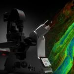
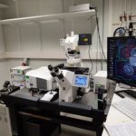
Photonic BioImaging is a Unit of Technology and Service (UTechS) providing optical imaging expertise in life sciences and especially their application in studies on infectious biology.

Our activities include service rendering, training, technology-driven research and technology development. They are highly multi-disciplined, and collaborative, with the mission goal focused on the use of quantitative imaging and analysis to understand the processes of cell/tissue-biology, and their usurpation by infection and disease. The R&D is founded upon the need to develop optical imaging methods that bring new understanding of host-pathogen interactions and in situ high-content imaging techniques and their application to infection, cell biology, cellular microbiology, and microbiology. We work on novel techniques extrapolating quantitative information on spatiotemporal dynamics in situ and we push the limits of existing approaches aiming to enhance their performance thereby broadening their experimental utility.

Practical Information

We are part of the C2RT (Center for Technological Resources and Research), a center gathering the Technology and Service units (UTechS) and the technological core facilities of the campus. As such we follow the common guidelines and good practices.

If you did not find what you were looking for, please check out the  DT Brochure

Instrument booking, training/project request :

PPMS Booking
(intranet access only)

More information : PBI_HOW TO REQUEST A TRAINING OR A PROJECT

Open Desk :

The Photonic BioImaging team (Imagopole) offers a bi-weekly virtual open-desk.The goal of the open-desk is to make the PBI engineers available for informal discussions about your forthcoming projects, or any imaging-related question you might have. There is no need to schedule an appointment, just come with your questions. Open-desks are planned to happen every two weeks on Teams on Mondays, (2:00pm to 3:30 pm). Please contact us (pbi.contact@pasteur.fr) for more information on the current schedules and login procedure.

Feedback / Comments

pbi.feedback@pasteur.fr

User Surveys Analysis

2018-Survey-UTechS PBI

2019 UTechS PBISurveyAnalysis

Equipments

Booking site
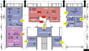
Show the location of equipments Hide location

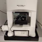
Label free imaging microscope • Nanolive • CX-A

NANOLIVE – CX-A Label-free imaging microscope The automated CX-A Nanolive’s is a structural label free microscope for live imaging. It generates holograms, by reporting the 3D refractive index (RI) distribution of all structures or […]

Microscope spinning disk • Andor • BC43

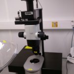
This system is an automated microscope equipped with two fluorescent imaging modalities: widefield and confocal (Borealis™-illuminated multipoint). This system is easy to use, offers different support, suitable for a wide variety of sample types […]

Microscope Opera Phenix Plus • Revvity • Opera Phenix Plus

The Opera Phenix Plus is an automated microlens-enhanced spinning disk confocal microscope, designed for high-throughput / high-content assays and phenotypic screening. Compatible with multiple carriers (from slides to 1536-well plates), it is mainly dedicated […]

Vaheat – Temperature control device • Interherence • Vaheat

Vaheat Heating system The Vaheat system is a device allowing control of the temperature directly under your sample. Few characteristics: Range of temperature : Room temperature up to 100°C . Precision : Relative temperature […]

Automated widefield/confocal microscope • ZEISS • CD7 - 900 Airyscan 2

This system is an automated microscope equipped with three major modalities : widefield, confocal and Airyscan. This methodological flexibility, combined with a wide range of analytical tools, means that we can work on different […]

Multiphoton microscope • Miltenyi Biotec • TrimScope Matrix

Our brand new multiphoton microscope : TrimScope Matrix Localization: 26-01-13D – BSL1 room Features: Lasers: Ti:Sapphire : Chameleon Ultra II, 80MHz, COHERENT (Excitation 680 nm to 1000 nm) O.P.O. : Insight X3 Dual, Spectra-physics    […]

Microscope confocal inverted FLIM • Leica • SP8

Inverted microscope fully motorized, equipped with a laser scanning confocal head and a TIRF unit. It allows the acquisition in spectral mode, multi-positions,  multi-mosaics. Features : Available light sources 405 nm 50 mW 488 […]

Microscope Super resolution SIM/SMLM • ZEISS • Elyra 7

This particular system is equipped with two of the major super resolution modalities Lattice SIM (Structured Illumination Microscopy) and 3D SMLM (Single Molecule Localisation Microscopy) allowing the observation of fixed and living biological phenomena […]

High Content Analysis • Perkin Elmer • Opera Phenix

System Characteristics Available Objectives 5x air (NA 0.16, WD 12.1mm, ~ 2.583 μm x 2.583 μm). 10x air (NA 0.3, WD 5.2mm ~ 1.292 μm x 1.292 μm). 20x air (NA 0.4, WD 8.28mm […]

Microscope spinning disk • Nikon • Ti2E - Yokagawa CSU W1

Spinning-disk Ti2E Inverted microscope, equipped with a Yokagawa CSU-W1 spinning disk with 2 pinhole size, and two cameras: a more sensitive and another more resolutive. It allows the fast acquisition of optically sectioned slices at […]

Light Sheet microscope • Miltenyi Biotech - Lavision Biotech • UltraMicroscope II

imaging in aqueous buffers and organic solvent  optical zoom from 1.26x to 12.6x  large sample uptake up to 30 mm x 30 mm x 15 mm  dynamic horizontal focusing for better Z resolution  6 […]

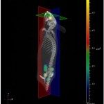
FMT 4000 Perkin Elmer • Perkin Elmer • FMT 4000

Small animal pre-clinical in vivo imaging system dedicated to 3D imaging of fluorescence signal (Fluorescence Molecular Tomography). The system is equipped with 4 lasers in the Near Infra Red and is calibrated for quantifying […]

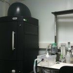
IVIS Spectrum CT Perkin Elmer • Perkin Elmer • IVIS Spectrum CT

Small animal pre-clinical in vivo imaging systems dedicated to 2D and 3D imaging of bioluminescence and fluorescence signals. The system is equipped with 10 excitation filters and 18 emission filters, and a high sensitive […]
IVIS Spectrum Perkin Elmer • Perkin Elmer • IVIS Spectrum

Small animal pre-clinical in vivo imaging systems dedicated to 2D and 3D imaging of bioluminescence and fluorescence signals. Each system is equipped with 10 excitation filters and 18 emission filters, and a high sensitive […]

CellVoyager CV1000 • Yokagawa • CV1000

Presentation. The Yokagawa CellVoyager CV1000 is an automated microscope based on the spinning-disk technology.    It excels at multi-scale microscopy, when locating rare events in a large sample is needed. Applications typically consist in […]

Microscope confocal SP5 HyD inverted • Leica • SP5 HyD resonnant scanner Matrix screener

Inverted microscope fully motorized, equipped with a laser scanning confocal head. It allows the acquisition in spectral mode, multi-positions,  multi-mosaics, and autofocus control with sensitive detectors.   Features : Available light sources 405 nm […]

Microscope confocal SP8 inverted • Leica • SP8

  Inverted microscope fully motorized, equipped with a laser scanning confocal head. It allows the acquisition in spectral mode, multi-positions,  multi-mosaics.   Features : Available light sources 405 nm White laser 470-670 nm Mercury […]

Microscope spinning-disk UltraView VOX • Perkin-Elmer • UltraView VOX

Spinning-disk UltraView VOX Inverted microscope, equipped with a Yokagawa CSUX1 spinning- disk, and two cameras to do dual-color simultaneous imaging. It allows the fast acquisition of optically sectioned slices at very high speed, and is […]

Microscope confocal LSM780 • ZEISS • LSM 780

Inverted confocal microscope (780 head) with a highly sensitive spectral detector GaAsp of 32 channels. Features :  Available laser lines for excitation Confocal 405 nm (30 mW) – Solid 458, 488, 512 nm (25 […]

Microscope widefield apotome inverted • ZEISS • ApoTome inverted

  Inverted widefield Microscope equipped with apotome grid allowing global fluorescence and optical sections imaging .   Features : Available light sources Mercury Lamp  HBO 100 Available filters Fluorescence Excitation Dichroic Emission DAPI BP […]

Microscope widefield apotome2 upright • ZEISS • ApoTome 2 upright

Upright widefield Microscope equipped with apotome2 grids allowing global fluorescence and optical sections imaging  and a color camera allowing the observation of histological slices.   Features : Available light sources Mercury Lamp  HXP 120 […]

Microscope widefield Definite Focus • ZEISS • Definite focus

Inverted widefield Microscope equipped with LED colibris  and mercury lamp allowing acquisition on live fluorescent samples with control of the focus using the ‘Definite Focus’ module.   Features : Available light sources Mercury Lamp […]

Microscope confocal LSM700 inverted • ZEISS • LSM700 inverted

  Inverted microscope, equipped with a laser scanning confocal head. It allows the acquisition of optically sectioned slices.   Features : Available light sources 405 nm 488 nm 555 nm 639 nm Halogen lamp […]

Microscope confocal LSM700 upright • ZEISS • LSM700 upright

  Upright microscope, equipped with a laser scanning confocal head. It allows the acquisition of optically sectioned slices.   Features : Available light sources 405 nm 488 nm 555 nm 639 nm Mercury lamp […]

Software

Members

Former Members

2000
2000
Name
Position
2017
2020
Anne-Marie Balazuc
Administrative Staff
2017
2020
Géraldine Bonnefoy
2017
2020
Julien Compagnon
Post-doc
2017
2020
Barbara Corelli
PhD Student
2017
2020
Debora Olivier (Keller)
Research Engineer
2017
2020
Jean-Yves Tinevez
Research Engineer
2017
2020
Sébastien Herbert
Research Engineer
2017
2020
Navnina Bhatia
Post-doc
2017
2020
Sophie Bouccara
Post-doc
2017
2020
Fabiane Sonego
Post-doc
2017
2020
Pascal Roux
Research Engineer
2017
2020
Malek Dimassi
Undergraduate Student
2017
2020
Malek Bouallegue
Undergraduate Student
2017
2020
Lesly Raulin
Research Engineer
2017
2020
Pierre Falanga
Permanent Researcher
2017
2020
Marie-Anne Nicola
Research Engineer
Ioanna Theodorou
Research Engineer
Myriam Ibilli
Administrative Staff
Christophe Gallard
Undergraduate Student
JonEun Ihm
Research Engineer
Show all

Courses

Principles and applications of fluorescence microscopy

This course is designed for Master’s and international PhD students planning to use advanced live-cell fluorescence microscopy in their future research. For more information and program, please, visit the course webpage. Program / Content […]

2025-03-10 09:00:00 2025-03-21 18:00:00 Europe/Paris Principles and applications of fluorescence microscopy This course is designed for Master’s and international PhD students planning to use advanced live-cell fluorescence microscopy in their future research. For more information and program, please, visit the course webpage. Program / Content […] Institut Pasteur, Rue du Docteur Roux, Paris, France

Microscopy – Principles and Applications of Fluorescence Microscopy

Fluorescence microscopy has revolutionized researchers’ ability to monitor cellular structures and dynamics. Over the last decades, the spectrum of applications of fluorescence microscopy has been constantly broadened by novel combinations of optics principles supported […]

2023-03-06 08:00:00 2023-03-17 19:00:00 Europe/Paris Microscopy – Principles and Applications of Fluorescence Microscopy Fluorescence microscopy has revolutionized researchers’ ability to monitor cellular structures and dynamics. Over the last decades, the spectrum of applications of fluorescence microscopy has been constantly broadened by novel combinations of optics principles supported […] 25-28 Rue du Dr Roux, 75015 Paris, France

Projects

Transversal Project

Fundings

Partners

Publications

Download

Contact

Phone:

+33(0)1 45 68 86 54 (N. Aulner, Core facility manager)

+33(0)1 40 61 39 14 (C. Da Agra, Assistant)

Email: PBI.contact@pasteur.fr

Address : 25-28 Rue du Docteur Roux 75015, Paris France