Search anything and hit enter
  • Teams
  • Members
  • Projects
  • Events
  • Calls
  • Jobs
  • publications
  • Software
  • Tools
  • Network
  • Equipment

A little guide for advanced search:

  • Tip 1. You can use quotes "" to search for an exact expression.
    Example: "cell division"
  • Tip 2. You can use + symbol to restrict results containing all words.
    Example: +cell +stem
  • Tip 3. You can use + and - symbols to force inclusion or exclusion of specific words.
    Example: +cell -stem
e.g. searching for members in projects tagged cancer
Search for
Count
IN
OUT
Content 1
  • member
  • team
  • department
  • center
  • program_project
  • nrc
  • whocc
  • project
  • software
  • tool
  • patent
  • Administrative Staff
  • Assistant Professor
  • Associate Professor
  • Clinical Research Assistant
  • Clinical Research Nurse
  • Clinician Researcher
  • Department Manager
  • Dual-education Student
  • Full Professor
  • Honorary Professor
  • Lab assistant
  • Master Student
  • Non-permanent Researcher
  • Nursing Staff
  • Permanent Researcher
  • Pharmacist
  • PhD Student
  • Physician
  • Post-doc
  • Prize
  • Project Manager
  • Research Associate
  • Research Engineer
  • Retired scientist
  • Technician
  • Undergraduate Student
  • Veterinary
  • Visiting Scientist
  • Deputy Director of Center
  • Deputy Director of Department
  • Deputy Director of National Reference Center
  • Deputy Head of Facility
  • Director of Center
  • Director of Department
  • Director of Institute
  • Director of National Reference Center
  • Group Leader
  • Head of Facility
  • Head of Operations
  • Head of Structure
  • Honorary President of the Departement
  • Labex Coordinator
Content 2
  • member
  • team
  • department
  • center
  • program_project
  • nrc
  • whocc
  • project
  • software
  • tool
  • patent
  • Administrative Staff
  • Assistant Professor
  • Associate Professor
  • Clinical Research Assistant
  • Clinical Research Nurse
  • Clinician Researcher
  • Department Manager
  • Dual-education Student
  • Full Professor
  • Honorary Professor
  • Lab assistant
  • Master Student
  • Non-permanent Researcher
  • Nursing Staff
  • Permanent Researcher
  • Pharmacist
  • PhD Student
  • Physician
  • Post-doc
  • Prize
  • Project Manager
  • Research Associate
  • Research Engineer
  • Retired scientist
  • Technician
  • Undergraduate Student
  • Veterinary
  • Visiting Scientist
  • Deputy Director of Center
  • Deputy Director of Department
  • Deputy Director of National Reference Center
  • Deputy Head of Facility
  • Director of Center
  • Director of Department
  • Director of Institute
  • Director of National Reference Center
  • Group Leader
  • Head of Facility
  • Head of Operations
  • Head of Structure
  • Honorary President of the Departement
  • Labex Coordinator
Search
Go back
Scroll to top
Share

About

Electron Microscopy at Ultrastructural BioImaging

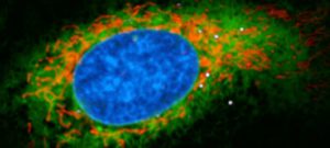
The Ultrastructural Bioimaging (UBI) facility at the Institut Pasteur provides state-of-the-art electron microscopy imaging approaches to users from the campus and outside. Samples range from single molecules to whole organisms. They can be observed at the highest resolution possible.
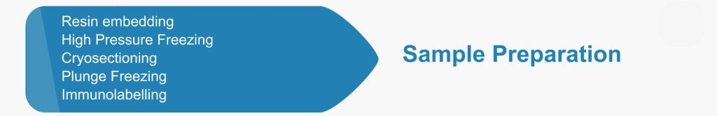
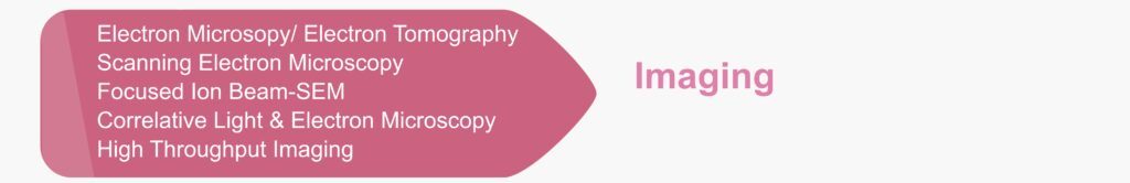
What we do:

Who are we?

13 electron microscopists with expertise covering a broad range of TEM & SEM methodologies.

What we offer:

  • Training
  • Collaboration or Service
  • Implementation & Development of new methods

How to get started:

  • Come and tell us more about your project in our weekly UBI team meeting or email us at ubi.all@pasteur.fr
  • New trainings / projects should be requested via our online system : PPMS

All samples with a biosafety level P2/P3 require the submission of a ‘protocol de recherche’ to document the procedure of inactivation.

Certifications and networks:

  • UBI is member of the Center for Technological Resources and Reearch (C2RT), centralizing the Technology and Service units (UTechS) and the technological core facilities of Institut Pasteur. Therefore, UBI follows the common C2RT guidlines for best working practices.
  • UBI is Ibisa labelled and ISO 9001 certified
  • UBI is part of FBI (France BioImaging), CTLS (Core Technologies for Life Sciences), C4L (Core for Life), Labex IBEID.

Brochure Technology Department C2RT-C2RA-C2RA-CRBIP

Localisations et guidelines :

DT-brochure-localisations

DT-brochure-guidelines

Job / internship : UBI-opportunities@pasteur.fr

Services

Booking site

UBI is a core facility with a broad mandate that supports a wide community of EM users. UBI offers services and hands-on training across various levels. Autonomous level users are able to book and use facility’s systems independently, while assisted users are followed closely by UBI members. On a basic service status, the facility staff  will prepare, observe samples and provide high quality images. In case of scientific collaborations UBI members will be actively involved to all required steps of the research project, including image processing. Collaborative projects are highly valued by UBI, as, along them, new methods can be implemented and developed in the facility.

Training in Electron Microscopy

UBI offers various hands-on trainings in Electron Microscopy covering a broad range of methods and techniques from sample preparation to imaging and image processing Negative staining (2 sessions x 3h) Sample preparation Ultramicrotomy (conventional […]

Image Processing for EM

  Tomographic reconstruction In electron tomography, the tilt series acquired, by tilting the sample to different angles under the electron beam (usually every 1 or 2 degrees from about −60° to +60°), can then […]

Correlative Light and Electron Microscopy

Correlative Light & Electron Microscopy (CLEM) approaches are methods of choice to image rare or dynamic events at high-resolution by Electron Microscopy (EM).  The functional and dynamic information coming from Light Microscopy (LM) can […]

Imaging

    Transmission electron microscopy In Transmission Electron Microscopy (TEM) the image is formed from the interaction of the electrons with the sample as the beam is transmitted through the specimen.  Transmission electron microscopes […]

Sample preparation

  Resin embedding The embedding of a soft biological sample in a plastic resin hardens it enough for thin sectioning. In this way the sample is ready for TEM, TEM-tomography, and FIB-SEM tomography. Classical […]

Single Particle Electron Microscopy

Negative stain and Cryo-electron microscopy is used to provide structural information of macromolecules and their complexes by imaging non-crystalline speciments (single particles) absorbed in electron microscopy grids. Fédry J, Liu Y, Péhau-Arnaudet G, Pei […]

Equipments

Booking site

High Pressure Freezing • Leica • EM ICE

Cryo-fixation has distinct advantages over chemical fixation. It is achieved instantly (within milliseconds) ensuring simultaneous immobilization of all macromolecules present. High Pressure Freezing Currently is the only method to vitrify thicker samples (up to […]

Image Processing Workstations for Electron Microscopy • •

All workstations can be booked through the PPMS system. Manet (named after Edouard Manet) Bi-processor Intel Xeon Nehalem 4c X5560 2.8GHz/ 80Go DDR3-1333 PNY NVIDIA GTX 1080 TI -11Go GDDR5X OS: CentOS Linux 7.7 […]

(Cryo-) Ultramicrotomes • Leica •

Ultramicrotomes allow easy preparation of semi- and ultra-fine sections of biological samples for Transmission EM or FIB-SEM observations. UBI is equipped with 3 ultramicrotomes for room temperature sectioning and 3 cryo-ultramicrotomes.  

Focused Ion Beam Scanning Electron Microscope • Zeiss • Auriga

  Focused ion beam scanning EM is typically applied to analyze whole cells in 3D. In 2016 Atlas-5 was installed on our Auriga with the goal to establish the automated imaging of serial sections, […]

Scanning Electron Microscope • Jeol • 6700F

SEM is typically applied to image the surface of samples. The Jeol 6700F is a routine SEM that can readily be handled by our users after training. It is also equipped with a cryo-transfer […]

Transmission Electron Microscope • FEI • Tecnai T12

The Ultrastructural BioImaging unit has two transmission EMs operated at 120kV with Lab6-electron source: T12 tomo and T12 cryo. T12 cryo is equipped with a cryo-box for prescreening of samples under cryogenic conditions. It […]

Transmission Electron Cryo Microscope • FEI • Tecnai F20

  The Tecnai F20 is a 200 kV Transmission Electron Microscope (TEM) specifically designed for high resolution data collection and tomography. The Field Emission Gun (FEG) provides high beam intensity and stability. Further structural […]

Automatic Plunge Freezer • Leica • EM GP

Plunge freezing For samples with thickness < few microns, such as isolated particles including proteins, macromolecular complexes, decorated liposomes, viruses, bacteria, eukaryotic cells…. Samples are deposited or grown directly onto EM grids and cryo-fixed […]

CryoCLEM Microscope • Leica • Leica EM Cryo CLEM

Correlative light and electron microscopy (CLEM) can provide localization and identification solutions of rare events/structures in crowded environments. Electron microscopy grids can be mounted on the Leica EM Cryo CLEM microscope. Full grid fluorescent […]

Members

Projects

Transversal Project

Fundings

Partners

Publications

Download

3D imaging

-Under construction-