
Please see the english version of the site :
https://research.pasteur.fr/en/team/virus-and-immunity/
https://research.pasteur.fr/en/team/virus-and-immunity/


2014-2015 – Céline Bressollette – Praticien Hospitalier
2007-2010 – Fabrizzio Mammano – Directeur de recherche Inserm
2001-2008 – Arnaud Moris – Directeur de recherche CNRS
2001-2009 – Nathalie Sol-Foulon – Chargée de recherche CNRS
POST-DOCTORANT
2020-2023 – Mathieu Hubert
2016-2021 – Blandine Monel
2018-2019 – Quentin Nevers
2015-2017 – Cécile Meunier
2015-2017 – Sonia Amraoui
2015-2017 – Nicolas Roy
2014-2014 – Nuno Miguel Ribeiro Palha
2013-2017 – Daniel Aaron Donahue
2013-2016 – Alex Compton
2013-2013 – Linda Obiang
2011-2016 – Jean-Jacques Mention
2010-2015 – Diana Ayinde
2010-2014 – Isabel Puigdomenech
2010-2011 – Eva Maria Weiss
2009-2010 – Stéphanie Louis
2009-2013 – Marine Malbec
2008-2010 – Samantha Brandler
2007-2012 – Jérôme Feldmann
2004-2006 – Fabien Blanchet
2003-2005 – Frédéric Delebecque
2002-2004 – Cécile Esnault
2001-2009 – Cinzia Nobile
PhD
2019-2022 – Ludivine Grezlak
2019-2022 – Jérôme Kervevan
2018-2021 – Maaran Michael Rajah
2018-2021 – Jérémy Dufloo
2013-2016 – Léa Richard
2013-2016 – Lise Chauveau
2010-2014 – Ferdinand Roesch
2010-2013 – Réjane Rua
2008-2010 – Marion Sourisseau
2007-2010 – Daniela Vendrame
2007-2011 – Alice Lepelley
2006-2009 – Dominika Rudnicka
Undergraduate Student
06/2023 – 08/2023 – Inès Maréchal
05/2023 – 02/2024 – Chloé Petiot
02/2023 – 08/2023 – Thibault Vanhoucke
04/2023 – 07/2023 – Anike Morch
2022-2023 – Augustin Martin
2022-2023 – Lou-Léna Vrignaud
2021-2022 – Cassandre Garnier
2020-2021 – Elodie Bishop
2017-2018 – Samy Sid Ahmed
2016-2017 – Guillaume Mestrallet
2015-2016 – Talya Raphael
2015-2015 – Louise Veron
2015-2016 – Norbert Couspel
2015-2015 – Anne Billet
2014-2015 – Anais Ducher
Assistante Administrative
Isma Ziani
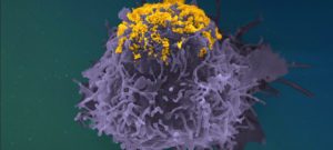
The MOOC offers a broad overview of the state of knowledge on viruses associated with cancer, the different mechanisms of carcinogenesis, the host immune response and progress in preventing tumors. Registration on: https://www.fun-mooc.fr/en/courses/viruses-and-human-cancers/ […]


Phone: +33 1 45 68 83 53 Email: olivier.schwartz@pasteur.fr Address Bât Lwoff 25-28 Rue du Docteur Roux 75015, Paris France