Tapez votre recherche ici
  • Équipes
  • Membres
  • Projets
  • Événements
  • Appels
  • Emplois
  • publications
  • Logiciel
  • Outils
  • Réseau
  • Équipement

Un petit guide pour l'utilisation de la recherche avancée :

  • Tip 1. Utilisez "" afin de chercher une expression exacte.
    Exemple : "division cellulaire"
  • Tip 2. Utilisez + afin de rendre obligatoire la présence d'un mot.
    Exemple : +cellule +stem
  • Tip 3. Utilisez + et - afin de forcer une inclusion ou exclusion d'un mot.
    Exemple : +cellule -stem
e.g. searching for members in projects tagged cancer
Rechercher
Compteur
IN
OUT
Contenu 1
  • member
  • team
  • department
  • center
  • program_project
  • nrc
  • whocc
  • project
  • software
  • tool
  • patent
  • Personnel Administratif
  • Chargé(e) de Recherche Expert
  • Directeur(trice) de Recherche
  • Assistant(e) de Recherche Clinique
  • Infirmier(e) de Recherche Clinique
  • Chercheur(euse) Clinicien(ne)
  • Manager de département
  • Etudiant(e) en alternance
  • Professeur(e)
  • Professeur Honoraire
  • Aide technique
  • Etudiant(e) M2
  • Chercheur(euse) Contractuel(le)
  • Personnel infirmier
  • Chercheur(euse) Permanent(e)
  • Pharmacien(ne)
  • Etudiant(e) en thèse
  • Médecin
  • Post-doctorant(e)
  • Prize
  • Chef(fe) de Projet
  • Chargé(e) de Recherche
  • Ingénieur(e) de Recherche
  • Chercheur(euse) Retraité(e)
  • Technicien(ne)
  • Etudiant(e)
  • Vétérinaire
  • Visiteur(euse) Scientifique
  • Directeur(trice) Adjoint(e) de Centre
  • Directeur(trice) Adjoint(e) de Départment
  • Directeur(trice) Adjoint(e) de Centre National de Référence
  • Directeur(trice) Adjoint(e) de Plateforme
  • Directeur(trice) de Centre
  • Directeur(trice) de Départment
  • Directeur(trice) d'Institut
  • Directeur(trice) de Centre National de Référence
  • Chef(fe) de Groupe
  • Responsable de Plateforme
  • Responsable opérationnel et administratif
  • Responsable de Structure
  • Président(e) d'honneur de Département
  • Coordinateur(trice) du Labex
Contenu 2
  • member
  • team
  • department
  • center
  • program_project
  • nrc
  • whocc
  • project
  • software
  • tool
  • patent
  • Personnel Administratif
  • Chargé(e) de Recherche Expert
  • Directeur(trice) de Recherche
  • Assistant(e) de Recherche Clinique
  • Infirmier(e) de Recherche Clinique
  • Chercheur(euse) Clinicien(ne)
  • Manager de département
  • Etudiant(e) en alternance
  • Professeur(e)
  • Professeur Honoraire
  • Aide technique
  • Etudiant(e) M2
  • Chercheur(euse) Contractuel(le)
  • Personnel infirmier
  • Chercheur(euse) Permanent(e)
  • Pharmacien(ne)
  • Etudiant(e) en thèse
  • Médecin
  • Post-doctorant(e)
  • Prize
  • Chef(fe) de Projet
  • Chargé(e) de Recherche
  • Ingénieur(e) de Recherche
  • Chercheur(euse) Retraité(e)
  • Technicien(ne)
  • Etudiant(e)
  • Vétérinaire
  • Visiteur(euse) Scientifique
  • Directeur(trice) Adjoint(e) de Centre
  • Directeur(trice) Adjoint(e) de Départment
  • Directeur(trice) Adjoint(e) de Centre National de Référence
  • Directeur(trice) Adjoint(e) de Plateforme
  • Directeur(trice) de Centre
  • Directeur(trice) de Départment
  • Directeur(trice) d'Institut
  • Directeur(trice) de Centre National de Référence
  • Chef(fe) de Groupe
  • Responsable de Plateforme
  • Responsable opérationnel et administratif
  • Responsable de Structure
  • Président(e) d'honneur de Département
  • Coordinateur(trice) du Labex
Recherche
Revenir
Haut de page
Partagez

Présentation

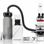
L’unité de BioImagerie Ultrastructurale offre à tous les groupes de recherche pasteuriens ainsi qu’aux groupes externes une aide scientifique et technologique en microscopie électronique. Nous proposons une grande diversité de préparations d’échantillons et de techniques d’imagerie et nous nous intéressons en particulier à la caractérisation de pathogènes et à leur interaction avec l’hôte. Notre équipe de spécialistes vous aidera à décider de la meilleure méthode à utiliser pour répondre à vos questions.

Les nouveaux projets doivent être soumis via notre système en ligne PPMS  : https://www.pasteur.eu/ppms/login/?pf=5

Il vous sera demandé de présenter brièvement votre question devant notre équipe.

Tous les échantillons présentant un risque de biosécurité niveau P2/P3 doivent être soumis à « un protocole de recherche » qui documente la procédure d’inactivation. L’acceptation de cette procédure peut prendre du temps dépendant de sa complexité.

L’unité de bio-imagerie ultra-structurale traite approximativement 100 projets par an. Nous encourageons les équipes qui ont de gros projets d’impliquer un personnel de son unité. Ils seront formés par les membres de l’unité.

Pour information, liens vers les brochures :

 Brochure Technology Department C2RT-C2RA-C2RA-CRBIP

Localisations et guidelines :

DT-brochure-localisations

DT-brochure-guidelines

contact pour candidature (job, stage) :  UBI-opportunities@pasteur.fr 

Services

Booking site

UBI est dotée d’un vaste  parc équipement qui lui permet de prendre en charge des formations ainsi que des projets de collaborations. UBI propose des services et des formations pratiques à différents niveaux. Les utilisateurs autonomes peuvent réserver et utiliser les systèmes de la plate-forme de manière indépendante, tandis que les utilisateurs assistés sont suivis de près par les membres de l’UBI. En collaboration scientifique, le personnel UBI préparera, observera des échantillons et fournira des images de haute qualité. Ils seront activement impliqués dans toutes les étapes requises du projet de recherche, y compris le traitement d’images. Les projets collaboratifs sont très appréciés par UBI, car, avec eux, de nouvelles méthodes peuvent être mises en œuvre et développées.

Training in Electron Microscopy

UBI offers various hands-on trainings in Electron Microscopy covering a broad range of methods and techniques from sample preparation to imaging and image processing Negative staining (2 sessions x 3h) Sample preparation Ultramicrotomy (conventional […]

Image Processing for EM

  Tomographic reconstruction In electron tomography, the tilt series acquired, by tilting the sample to different angles under the electron beam (usually every 1 or 2 degrees from about −60° to +60°), can then […]

Correlative Light and Electron Microscopy

Correlative Light & Electron Microscopy (CLEM) approaches are methods of choice to image rare or dynamic events at high-resolution by Electron Microscopy (EM).  The functional and dynamic information coming from Light Microscopy (LM) can […]

Imaging

    Transmission electron microscopy In Transmission Electron Microscopy (TEM) the image is formed from the interaction of the electrons with the sample as the beam is transmitted through the specimen.  Transmission electron microscopes […]

Sample preparation

  Resin embedding The embedding of a soft biological sample in a plastic resin hardens it enough for thin sectioning. In this way the sample is ready for TEM, TEM-tomography, and FIB-SEM tomography. Classical […]

Single Particle Electron Microscopy

Negative stain and Cryo-electron microscopy is used to provide structural information of macromolecules and their complexes by imaging non-crystalline speciments (single particles) absorbed in electron microscopy grids. Fédry J, Liu Y, Péhau-Arnaudet G, Pei […]

Équipements

Booking site

High Pressure Freezing • Leica • EM ICE

Cryo-fixation has distinct advantages over chemical fixation. It is achieved instantly (within milliseconds) ensuring simultaneous immobilization of all macromolecules present. High Pressure Freezing Currently is the only method to vitrify thicker samples (up to […]

Image Processing Workstations for Electron Microscopy • •

All workstations can be booked through the PPMS system. Manet (named after Edouard Manet) Bi-processor Intel Xeon Nehalem 4c X5560 2.8GHz/ 80Go DDR3-1333 PNY NVIDIA GTX 1080 TI -11Go GDDR5X OS: CentOS Linux 7.7 […]

(Cryo-) Ultramicrotomes • Leica •

Ultramicrotomes allow easy preparation of semi- and ultra-fine sections of biological samples for Transmission EM or FIB-SEM observations. UBI is equipped with 3 ultramicrotomes for room temperature sectioning and 3 cryo-ultramicrotomes.  

Focused Ion Beam Scanning Electron Microscope • Zeiss • Auriga

  Focused ion beam scanning EM is typically applied to analyze whole cells in 3D. In 2016 Atlas-5 was installed on our Auriga with the goal to establish the automated imaging of serial sections, […]

Scanning Electron Microscope • Jeol • 6700F

SEM is typically applied to image the surface of samples. The Jeol 6700F is a routine SEM that can readily be handled by our users after training. It is also equipped with a cryo-transfer […]

Transmission Electron Microscope • FEI • Tecnai T12

The Ultrastructural BioImaging unit has two transmission EMs operated at 120kV with Lab6-electron source: T12 tomo and T12 cryo. T12 cryo is equipped with a cryo-box for prescreening of samples under cryogenic conditions. It […]

Transmission Electron Cryo Microscope • FEI • Tecnai F20

  The Tecnai F20 is a 200 kV Transmission Electron Microscope (TEM) specifically designed for high resolution data collection and tomography. The Field Emission Gun (FEG) provides high beam intensity and stability. Further structural […]

Automatic Plunge Freezer • Leica • EM GP

Plunge freezing For samples with thickness < few microns, such as isolated particles including proteins, macromolecular complexes, decorated liposomes, viruses, bacteria, eukaryotic cells…. Samples are deposited or grown directly onto EM grids and cryo-fixed […]

CryoCLEM Microscope • Leica • Leica EM Cryo CLEM

Correlative light and electron microscopy (CLEM) can provide localization and identification solutions of rare events/structures in crowded environments. Electron microscopy grids can be mounted on the Leica EM Cryo CLEM microscope. Full grid fluorescent […]

Membres

Projets

Projet Transversal

Financements

Partenaires

Publications

Télécharger

BioImagerie Ultrastructurale

-Under construction-