Search anything and hit enter
  • Teams
  • Members
  • Projects
  • Events
  • Calls
  • Jobs
  • publications
  • Software
  • Tools
  • Network
  • Equipment

A little guide for advanced search:

  • Tip 1. You can use quotes "" to search for an exact expression.
    Example: "cell division"
  • Tip 2. You can use + symbol to restrict results containing all words.
    Example: +cell +stem
  • Tip 3. You can use + and - symbols to force inclusion or exclusion of specific words.
    Example: +cell -stem
e.g. searching for members in projects tagged cancer
Search for
Count
IN
OUT
Content 1
  • member
  • team
  • department
  • center
  • program_project
  • nrc
  • whocc
  • project
  • software
  • tool
  • patent
  • Administrative Staff
  • Assistant Professor
  • Associate Professor
  • Clinical Research Assistant
  • Clinical Research Nurse
  • Clinician Researcher
  • Department Manager
  • Dual-education Student
  • Full Professor
  • Honorary Professor
  • Lab assistant
  • Master Student
  • Non-permanent Researcher
  • Nursing Staff
  • Permanent Researcher
  • Pharmacist
  • PhD Student
  • Physician
  • Post-doc
  • Prize
  • Project Manager
  • Research Associate
  • Research Engineer
  • Retired scientist
  • Technician
  • Undergraduate Student
  • Veterinary
  • Visiting Scientist
  • Deputy Director of Center
  • Deputy Director of Department
  • Deputy Director of National Reference Center
  • Deputy Head of Facility
  • Director of Center
  • Director of Department
  • Director of Institute
  • Director of National Reference Center
  • Group Leader
  • Head of Facility
  • Head of Operations
  • Head of Structure
  • Honorary President of the Departement
  • Labex Coordinator
Content 2
  • member
  • team
  • department
  • center
  • program_project
  • nrc
  • whocc
  • project
  • software
  • tool
  • patent
  • Administrative Staff
  • Assistant Professor
  • Associate Professor
  • Clinical Research Assistant
  • Clinical Research Nurse
  • Clinician Researcher
  • Department Manager
  • Dual-education Student
  • Full Professor
  • Honorary Professor
  • Lab assistant
  • Master Student
  • Non-permanent Researcher
  • Nursing Staff
  • Permanent Researcher
  • Pharmacist
  • PhD Student
  • Physician
  • Post-doc
  • Prize
  • Project Manager
  • Research Associate
  • Research Engineer
  • Retired scientist
  • Technician
  • Undergraduate Student
  • Veterinary
  • Visiting Scientist
  • Deputy Director of Center
  • Deputy Director of Department
  • Deputy Director of National Reference Center
  • Deputy Head of Facility
  • Director of Center
  • Director of Department
  • Director of Institute
  • Director of National Reference Center
  • Group Leader
  • Head of Facility
  • Head of Operations
  • Head of Structure
  • Honorary President of the Departement
  • Labex Coordinator
Search

← Go to Research

Go back
Scroll to top
Share
© K. Melican.
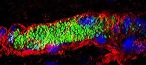
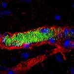
Human microvessel (red) colonized by N. meningitidis (green).

About

A number of infectious agents, including emerging pathogens and agents responsible for nosocomial infections, reach the blood during infection leading to septicemia and meningitis. Despite antibiotic therapy these infections result in severe sequelae and high death rates. A better understanding of the mechanisms of disease is a necessary step for the identification of innovative treatments.

We study the pathogenesis of Neisseria meningitidis (or meningococcus), a Gram-negative bacterium that recapitulates these different pathological effects. This bacterium asymptomatically colonizes the human nasopharynx and pathology is initiated when the bacterium crosses the nasopharynx epithelium and reaches the bloodstream where they survive and proliferate.
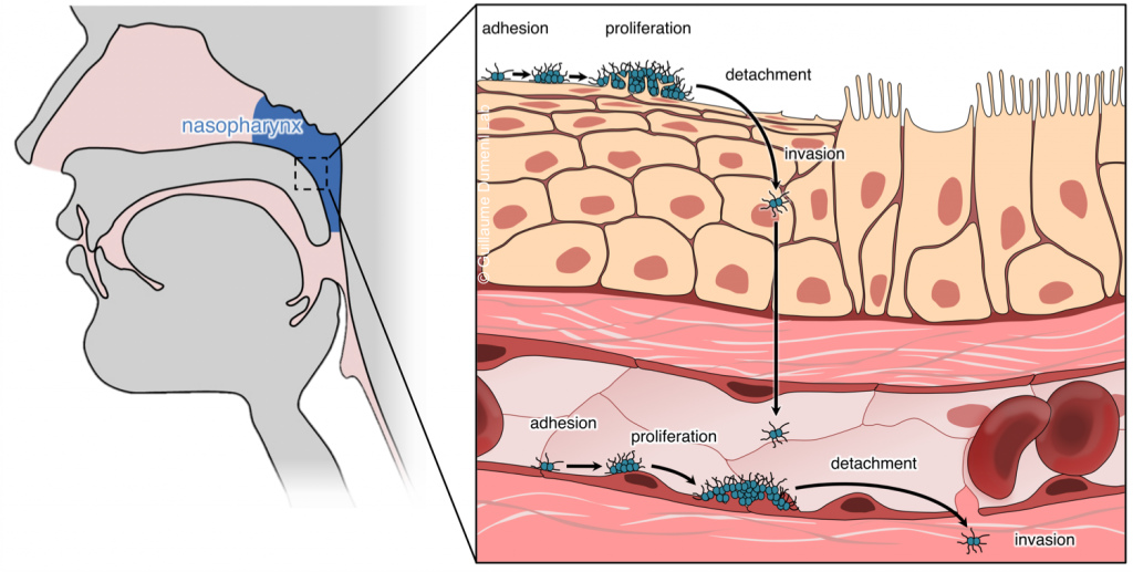
Carriage and pathogenesis of N. meningitidis

Outstanding questions in terms of understanding N. meningitidis pathogenesis include: how do bacteria cross the epithelium and reach the bloodstream? How do they survive in the blood? How do they damage vessels and reach the cerebrospinal fluid (i.e. cause septic shock and meningitis)? These questions open broader studies regarding tissue biology, bacterial adaptation to different environments and basic functions of the innate immune system.   We address these questions at different scales.

  •  Structural biology of bacterial virulence factors. We study in particular type IV pili and the molecular machinery necessary for their dynamic assembly and disassembly which allow bacteria to adhere host cells and are essential for virulence. We take advantage of single particle Cryo-Electron Microscopy and Cryo-Electron Tomography approaches.
  • Interaction of bacteria with host cellsNeisseria spp. are extracellular bacteria which manipulate host cell functions from the outside. We study how they mediate this intricate cross-talk with human endothelial cells in culture using fluorescence video microscopy and Cryo-Electron Tomography. We use 2D cultures and 3D microfluidics-based vessel-on-a-chip set-ups to study the impact at the tissue level. This leads us to basic cell biology and mechanobiology questions.
  • Impact of infection on blood vessel function. We have developed an animal model based on the xenograft of human skin onto immunodeficient mice. Because of its strict human specificity N. meningitidis type IV pili bind only to capillaries present in the human tissue.  Availability of such a model of infection recapitulating the cardinal features of the blood phase of the infection now allows us to address key questions in terms of the bacterial and host processes involved in N. meningitidis-caused vascular damage.
  • Innate immune system. Our humanized animal model also allows us to address how the innate immune system reacts to the intravascular infections and also how bacteria evade the immune system.

We thus follow a global and multidisciplinary approach combining microbiology with cell biology, vascular biology, chemistry and physics to study the pathogenesis of N. meningitidis infection.

Whenever possible our basic findings are exploited to design preventive and therapeutic approaches in collaboration with pharmacological companies. It is also essential to us to apply our understanding of N. meningitidis to the study of other bacterial pathogens involved in similar pathologies.

 

Former Members

Projects

Transversal Project

Fundings

Publications

Download

Alumni

Ximing Xu (Post-doctoral fellow 2014-2016) Maëlys Loh (Summer student 2016) Corinne Millien (Technician 2010-2015) Flore Aubey (Engineer 2013-2015) Anne-Flore Imhaus (PhD student 2009-2014) Silke Machata (Post-doctoral fellow 2010-2014) Valentina Lo Schiavo (Post-doctoral fellow 2012-2014) Idonya Aghoghogbe (M2 student 2014) Keira Melican (Post-doctoral fellow 2011-2013) Magali Soyer (PhD student 2009-2012) Paula Michea-Veloso (PhD student 2009-2012) Tiffany Martin (M2 student 2011-2012) Audrey Dumont (Post-doctoral fellow 2009-2010) Guillain Mikaty (PhD student 2007-2010) Benoît Rousseau (M2 student 2003-2004) Fanny Lanternier (M2 student 2003-2004) Emilie Mairey (PhD student 2004-2007)

Lab news

  • August 2021 Daria Bonazzi awarded an ANRJCJC funding
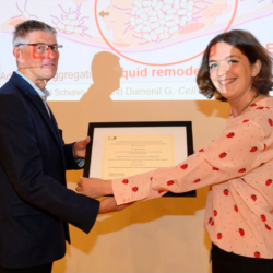
  • June 2021 Guillaume Duménil receives the Grand prix Fondation NRJInstitut de France
  • October 2019 Creation of the ” Pathogenesis of Vascular Infections ” INSERM unit
  • June 2019 Guillaume Duménil receives the Pasteur Vallery Radot prize Daria Bonazzi receives the Institut Pasteur young fellowship award
  • October 2016 Daria Bonazzi selected among the 10 finalists of the 2016 ASCB Kaluza Prize Daria Bonazzi recipient of a L’Oréal-UNESCO For Women in Science Fellowship

Pictures & Media

  • Broad public diffusion of knowledge
    • La bacteria Jekyll y Hyde – Guillaume Dumenil y Virginia Garretón,
      Puerto de Ideas Antofagasta Science Festival 2022

    • The Duménil lab was selected to produce a CellDance video for the ASCB-EMBO 2017 conference that took place in Philadelphia in December. Check it out!

    • Conference by Guillaume Duménil on the Vascular Colonization by N.meningitidis:

    Research videos
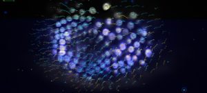
    • Adhesion of meningococcus aggregates to endothelial cells leads to ezrin protein accumulation (which represents plasma membrane remodelig). Ezrin-GFP transfected cells (in green) were infected by mCherry-expressing meningococcus aggregates (in red). Behaviour of ezrin was monitored over a 10 min period. As the aggregates reach the cells, ezrin started to accumulate. The scale bar corresponds to 20 μm. Author: Arthur Charles-Orszag Related to the article Soyer et al., Cell. Microbiol. 2014:

    • Meningococcus growth as a microcolony is accompanied by ezrin protein accumulation (which represents plasma membrane remodelig). Ezrin-GFP transfected cells (green) were infected by individual mCherry-expressing meningococci (red). Following initial adhesion (white arrows), behaviour of ezrin underneath the nascent microcolony was monitored over a 4 h period under flow conditions. As the microcolony reached a size of about 10 diplococci (30 min), ezrin started to accumulate and the cortical plaque size increased with the size of the microcolony. The scale bar corresponds to 20 μm. Author: Magali Soyer From the article Soyer et al., Cell Microbiol 2014.

    • Intravital microscopy showing a human vessel in the base of the skin graft labeled with UEA lectin (red) and perfused by the mouse circulation. Blood plasma is labeled with a 150 kDa FITC-dextran (green) introduced intravenously. Moving black silhouettes within the plasma are blood cells. Supplementary movie from Melican et al., PLOS Pathogens 2013.

  • Intravital microscopy showing adhesion of N. meningitidis (GFP, green) to a human vessel (UEA lectin, red) grafted onto an immunodeficient mouse, 30 min post infection. Supplementary movie from Melican et al., PLOS Pathogens 2013.

Contact

Adresse: Institut Pasteur – Unité Pathogénèse des infections vasculaires-28 rue du Docteur Roux – 75015 Paris