PhD position:
PhD position available to study the hearing system at the nanoscale using cutting-edge cryo-electron tomography & super-resolution microscopy? Join us with the Institut Pasteur Paris University International Doctoral Program (PPU) !!! bit.ly/3zbynLJ
https://twitter.com/RuppSartori/status/1584867862996742145?s=20&t=CUnah-3etCSHlTfl1c3bOA
PROFILE
- Expert Research Engineer
- PhD in Physics
- Expertise: Cryo-electron tomography, Cryo-correlative light and electron microscopy (cryoCLEM) & Image analysis applied to the study of eukaryotic cells, bacteria, viruses and host-pathogen interactions
- Research interests: Technological and methodological development for designing novel 3D cellular cryo-CLEM pipelines with super-resolution light microscopy, applied to single and multicellular systems.
- 03/2022-… Expert Research Engineer, NanoImaging Core Facility, Institut Pasteur, Paris, FR
- 2007-2022 Research Engineer, Ultrastructural BioImaging Unit, Institut Pasteur, Paris, FR
- 2014-2016 Visiting Scientist, LBME lab, Toulouse, FR
- 2004-2007 Postdoctoral fellow, MPI of Biochemistry, Munich, DE
- 2001-2004 Postdoctoral fellow, Institut Curie UMR168, Paris, France
- 1998- 2001 PhD in Mathematical Physics, Imperial College, London, UK
- 1995- 1996 Erasmus student, Imperial College, London, UK
- 1991- 1997 Master Degree in Physics, University of Padua, Italy
________________________________________________________________________________
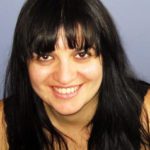
Anna is a Physicist with a strong passion for Imaging and Microscopy. After obtaining a degree in Physics at the University of Padua, Italy, and a PhD in Physics at Imperial College, London, UK, she turned towards the world of Microscopy in Life Science. She was granted a Postdoctoral Marie Curie Individual Fellowship at Institut Curie, Paris, where she focused on fluorescent videomicroscopy to model DNA migration in polymeric matrices. Then, she moved on for a second PostDoc in the cryo-EM renowned lab of Prof. W. Baumeister at the MPI of Biochemistry in Munich, where she established a novel technique, the correlation between fluorescent microscopy in cryogenic conditions and cryo-electron tomography (cryo-CLEM).
Since 2007, she works at the UBI platform as an expert of 3D cryo-electron microscopy. She coordinates collaborative scientific projects involving high resolution cryo-tomography and cryo-CLEM applied to the study of host-pathogen interaction, starting from sample preparation to data collection and analysis. In one of her latest research works in collaboration with the group of Prof. C. Zurzolo at Institut Pasteur, she conceived a novel cryo-CLEM approach that uncovered for the first time the striking actin’ structural organization of Tunneling Nanotubes in neuronal cells.
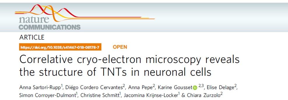
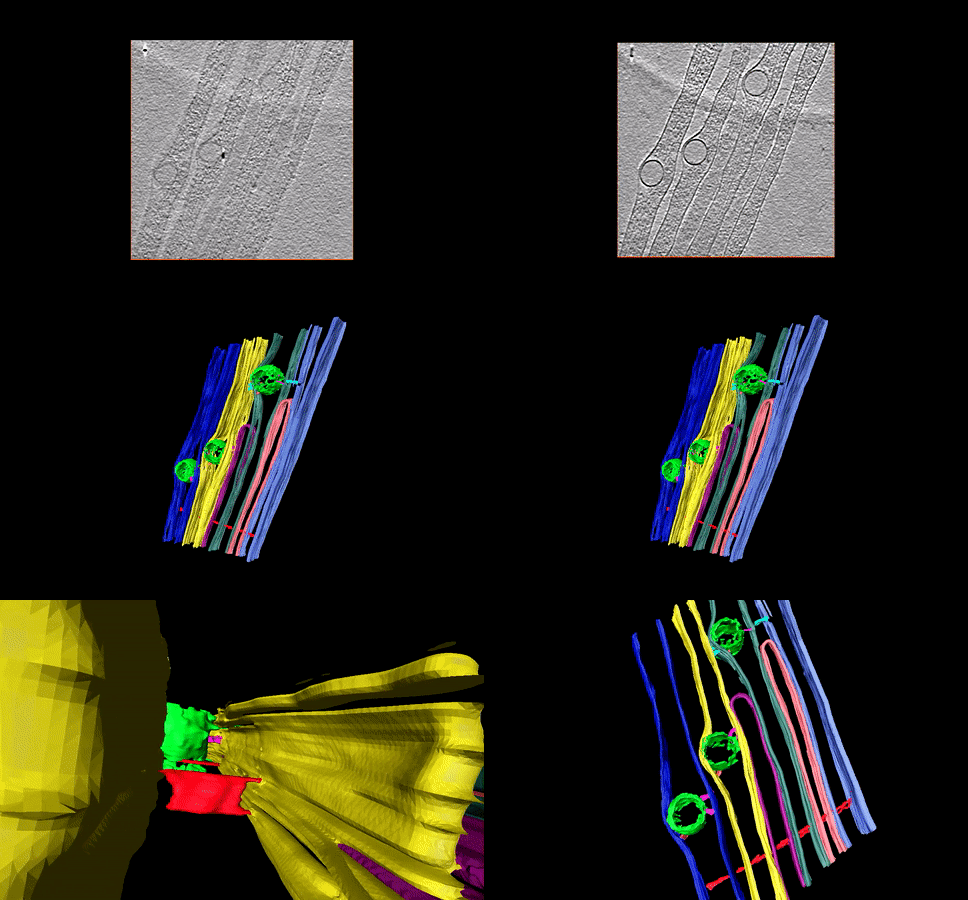 Cryo-electron tomogram reconstruction of Tunnelling Nanotubes (TNTs) connecting neuronal cells composed of individual TNTs (iTNTs) running parallel to each other and containing actin parallel bundles and cellular compartments. 3D rendering showing transport of vesicles (green) linked to the iTNTs plasma membrane (pink), linkers between the iTNTs (cyan) & threads connecting individual iTNTs (red). https://www.youtube.com/watch?v=kKcwm1AHZ9g
She is very fond of technological/methodological development, in particular of designing novel workflows for 3D cryo-CLEM of cellular systems, including cryo-focused ion beam (cryo-FIB) and cryo-lamellae. She is also actively testing and implementing cutting edge technologies in partnership with companies (e.g. Leica).
……………………………………………………………………………………………………………………………………………………….
Since the advent of the NanoImaging Core facility (NICF) at Institut Pasteur, featuring among the most advanced cryo-EM technologies available worldwide, including a Titan Krios with a Falcon 4i direct electron detector, a SelectrisX imaging filter and Phase plates, an Aquilos2 cryo-FIB/SEM for cryo-lamellae milling with iFLM module, she has set up advanced cryo-CLEM pipelines aimed at the study of host-pathogen interactions in collaboration, with Léa Swistak in the group of Jost Enninga and with Stéphane Tachon from the NICF.
She setup cryo-tomography at the UBI and NICF core facilities and introduced the technique to the campus.
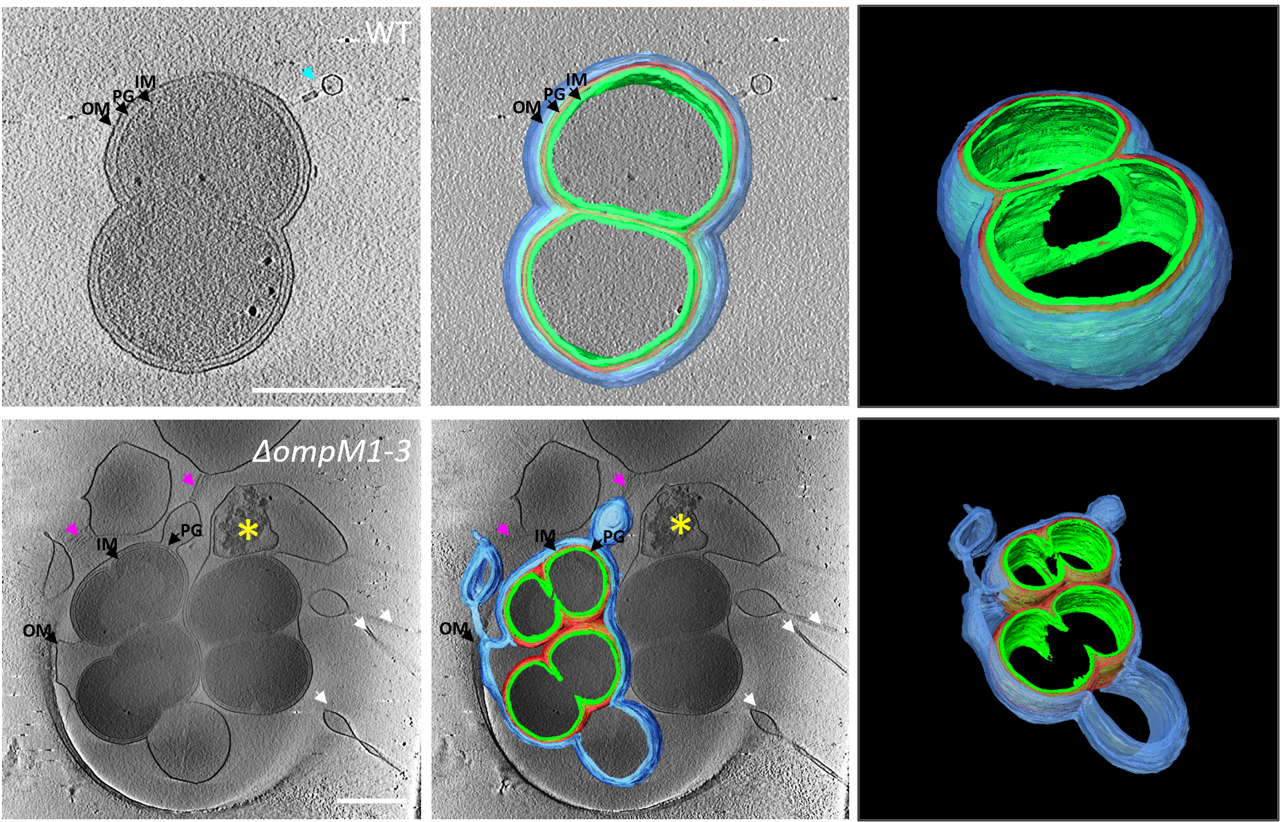
Cell envelope mutants in Veillonella parvula, “An ancient divide in outer membrane tethering systems in bacteria suggests a mechanism for the diderm-to-monoderm transition”. Witwinowski, J., Sartori-Rupp, A., Taib, N., Pende, N., Tham, N., Poppleton, D., Ghigo, J.-M., Beloin, C. & S. Gribaldo. Nat. Microbiol. 7:411-22. https://doi.org/10.1038/s41564-022-01066-3
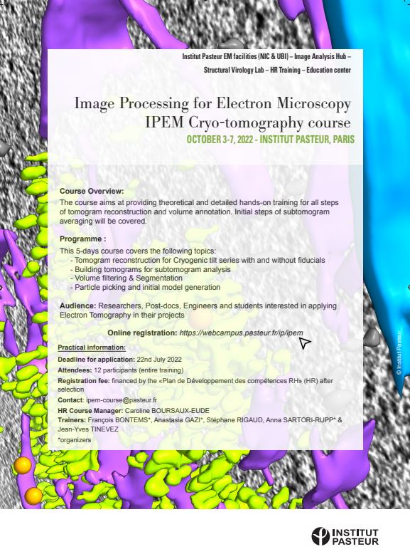
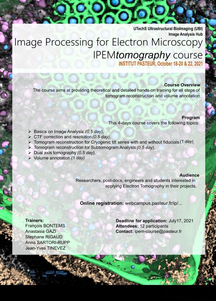
In October 2021 and October 2022 she organised together with Anastasia Gazi, Francois Bontems and Jean-Yves Tinevez the first and second image processing for electron microscopy (IPEMtomo) courses at Institut Pasteur.
