The brain senses and adapts to a wide range of environmental changes throughout life. We are interested in understanding how the neural stem cells and their cellular microenvironment (especially the niche cells) are able to detect and integrate different types of local and environnmental stimuli, from localised cellular growth to infection from neurotropic pathogens. We are relying on the powerful genetics of Drosophila as a model system, as well as on state-of-the-art transcriptional profiling and imaging to unravel the underlying mechansims of this adaptation. This will lead to a better picture of cellular interactions and dynamics in the brain under stress.
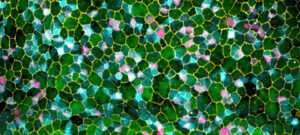
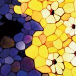
OMX-acquired image of a Drosophila blood-brain barrier (projection, early larval stage).
Green, Septate junctions.
Red, gap junction (Innexin 1).
Blue, Laminin.
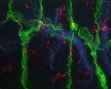
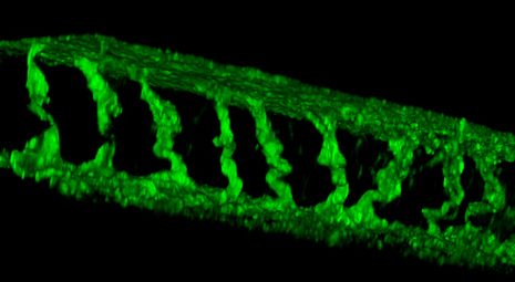
Confocal image showing a lateral view of a Drosophila ventral nerve chord (projection, early larval stage).
Green, neural stem cell membranes.
Red, neurons.
Blue, glia.
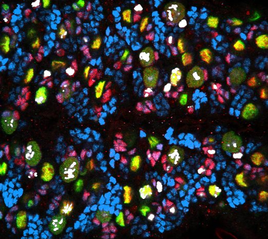
Confocal image of a ventral view of a Drosophila ventral nerve chord (single slice, late larval stage).
Green, neural stem cells.
Red, fate determinant Prospero.
Blue, neurons.
White, phospho-histone 3.
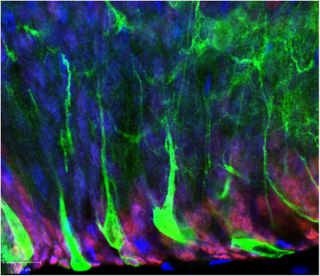
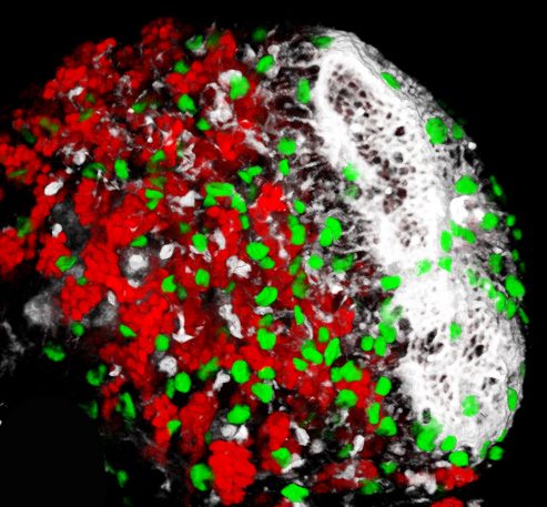
Tri-dimensional reconstruction of a confocal image showing Drosophila central brain and optic lobe (stack, late larval stage).
Green, glia.
Red, neurons.
White, actin.
[Improvision Data]
ImageName=Snapshot of 1.oif copy
TimeStampMicroSeconds=3515060629731706
TimeStamp=15:43:49,731 on 21 May 2015
ChannelName=
ChannelNo=1
TimepointName=1
TimepointNo=1
ZPlane=1
BlackPoint=0
WhitePoint=255
WhiteColour=255,255,255
XCalibrationMicrons=1
YCalibrationMicrons=1
ZCalibrationMicrons=1
TotalChannels=1
TotalTimepoints=1
TotalZPlanes=1
Software=Volocity 6.3.0
SampleUUID=c7daf462-b472-4dfc-b254-aed4d1481b82
Tri-dimensional reconstruction of a confocal image showing a Drosophila blood-brain barrier (stack, lateral view, early larval stage).
Green, blood-brain barrier membrane.