Viruses were for quite long characterized as the invisible heteronomous agents, (unseen by light microscopy & unable to propagate themselves in the absence of susceptible cells) capable of escaping the bacteriological filters (Rivers, 1932, The nature of viruses).
It was the birth and rise of the Electron Microscope that finally revealed the true nature of viruses, giving an exponential boost to Virology: Viruses are indeed so small that enter the susceptible cells and hijack their cellular machineries to replicate their genomes and produce new virions. The exact strategy adopted by each one of them is witnessed beautifully in the outstanding remodeling of the host cellular architecture during their life cycle.
While Single Particle reconstruction methods revealed the beauty of amazing symmetrical capsids in homogeneous samples, heterogeneous samples such as pleomorphic viruses, could only be studied by Electron Tomography (ET). The heterogeneous environment of the host and its remodeling takes place also in three dimensions. With the 3rd spatial aspect lost in 2D projection images, ET will always remain the method of choice to study their life cycles (attachment, entry, replication, assembly and egress) in molecular detail.
Highlights
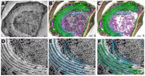
Archaeal viruses can surprise you! The lack of evolutionary relationship to other known viruses points to unique viral-host interaction sceneries. Sulfolobus islandicus filamentous virus (SIFV) is only one of them! In eukaryotes usually acquire envelopes inside the cell by budding through organelles. How is this taking place in Archaea that lack such organelles? SIFV was found to reach its full maturity (2 μm in length) and to be enveloped inside its host!
 PNAS 2021 Vol. 118 No. 32 e2105540118
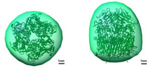
Newly synthesized Vaccinia membranes inside the eukaryotic host cell appear as short, curved units, named crescents. These develop into spheres where the viral DNA is packed. This step is critically depended on the viral scaffold protein D13. The proper assembly of the D13 scaffold layer around the convex side of the crescents and subsequently around the spheres is further fine tuned with the help of the Virus Membrane Associated Proteins (VMAPs). When the expression of the VMAP H7 is blocked, the D13 trimers instead of packing in the honeycomb lattice were found clustered together with short membranes of probable ER origin.

