Our research group focuses on the means of controlling chronic viral infections. We aim to reconstruct human tissues in organ-on-chips, to directly evaluate the capacity of antiviral immune cells to reach and target infected cells within tissues. We are also harnessing single cell approaches to define optimal antiviral T cells that could be amplified for immunotherapeutic approaches and then tested in organ-on-chip models. Building on these technologies, we aim to characterize the type of immune responses that can efficiently control viruses in lymphoid tissue and the respiratory mucosa.
Research Axis 1: Development of a lymphoid organ-on-chip to evaluate candidate antiviral vaccines
Organ-on-chips represent 3D cultures of primary human cells that are continuously perfused with nutrient medium by a microfluidics system. Human cells are provided with a more physiological environment than in classical 2D cultures and self-organize to reconstitute human tissues. Organs-on-chips provide new models to better mimic human physiology and help predict the effects of antiviral strategies.
We have developed a two-compartment lymphoid organ-on-chip (LO chip) based on microfluidics technology. We found that the LO chip provides a highly sensitive system to measure the interactions between antiviral CD4+ T cells and memory B cells from volunteers who received a COVID vaccine (R. Jeger-Madiot et al., J Exp Med 2024). The LO chip will be used to compare different vaccine boosting strategies directed at containing respiratory viruses and will be tested for its capacity to predict antigen immunogenicity.
The LO chip will also be used to evaluate the mechanisms underlying efficient antiviral responses in natural HIV Controllers. We have previously shown that HIV Controllers are characterized by particularly efficient CD4+ T cell responses associated with high TCR affinities against HIV Gag epitopes (M. Galperin et al., Sci. Immunol 2018; M. Claireaux et al., Nat Com 2022). We will determine the impact of these high affinity CD4+ T cells on viral dissemination and B cell memory responses within the LO chip model.
Research Axis 2: Development of airway and lung-on-chip models to decipher coronavirus pathogenesis
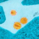
We are also using reconstructed airway epithelia to study the persistence of SARS-CoV-2 in its primary targets, the ciliated cells. We showed in this system that SARS-CoV-2 induces a loss of motile cilia in its target cells, which impairs the clearance of viral particles and may facilitate viral dissemination (R. Robinot, Nat Comm 2021). The image below shows a partially deciliated cells (green) with SARS-CoV-2 particles accumulating close to the plasma membrane (cyan).
We will further develop this system to generate an airway organ-on-chip that also includes a perfused vascular compartment. This system will be used to dissect the determinants of SARS-CoV-2 tropism and explore possible mechanisms of viral persistence. The airway-on-chip will also be key to evaluate the capacity of TCR-transduced T cells cells to migrate into the epithelial layer and target SARS-CoV-2 infected cells. The project is funded by 3D-LUNGO PEPR and is done in close collaboration with the team the Biomaterials and Microfluidics core facility of the Pasteur Institute.
Research Axis 3: Understanding the alteration of antiviral responses in Long COVID
Long COVID is recognized a major public health problem, as more than 10% of patients with SARS-CoV-2 infection show persisting symptoms more than 3 months after the acute infection stage. Long COVID patients suffer from a variety of persisting symptoms, in which debilitating fatigue, brain fog, and dyspnea predominate. The causes of this post-viral syndrome remain mostly unknown. In the PERSICOT ANRS project, we are testing the hypothesis that an inefficient T cell response may allow the SARS-CoV-2 virus to persist at low level and cause inflammatory damage in Long COVID patients.