Search anything and hit enter
  • Teams
  • Members
  • Projects
  • Events
  • Calls
  • Jobs
  • publications
  • Software
  • Tools
  • Network
  • Equipment

A little guide for advanced search:

  • Tip 1. You can use quotes "" to search for an exact expression.
    Example: "cell division"
  • Tip 2. You can use + symbol to restrict results containing all words.
    Example: +cell +stem
  • Tip 3. You can use + and - symbols to force inclusion or exclusion of specific words.
    Example: +cell -stem
e.g. searching for members in projects tagged cancer
Search for
Count
IN
OUT
Content 1
  • member
  • team
  • department
  • center
  • program_project
  • nrc
  • whocc
  • project
  • software
  • tool
  • patent
  • Administrative Staff
  • Assistant Professor
  • Associate Professor
  • Clinical Research Assistant
  • Clinical Research Nurse
  • Clinician Researcher
  • Department Manager
  • Dual-education Student
  • Full Professor
  • Honorary Professor
  • Lab assistant
  • Master Student
  • MD-PhD Student
  • Medical Staff
  • Non-permanent Researcher
  • Nursing Staff
  • Permanent Researcher
  • Pharmacist
  • PhD Student
  • Physician
  • Post-doc
  • Prize
  • Project Manager
  • Research Associate
  • Research Engineer
  • Retired scientist
  • Technician
  • Undergraduate Student
  • Veterinary
  • Visiting Scientist
  • Deputy Director of Center
  • Deputy Director of Department
  • Deputy Director of National Reference Center
  • Deputy Head of Facility
  • Director of Center
  • Director of Department
  • Director of Institute
  • Director of National Reference Center
  • Group Leader
  • Head of Facility
  • Head of Operations
  • Head of Structure
  • Honorary President of the Departement
  • Labex Coordinator
Content 2
  • member
  • team
  • department
  • center
  • program_project
  • nrc
  • whocc
  • project
  • software
  • tool
  • patent
  • Administrative Staff
  • Assistant Professor
  • Associate Professor
  • Clinical Research Assistant
  • Clinical Research Nurse
  • Clinician Researcher
  • Department Manager
  • Dual-education Student
  • Full Professor
  • Honorary Professor
  • Lab assistant
  • Master Student
  • MD-PhD Student
  • Medical Staff
  • Non-permanent Researcher
  • Nursing Staff
  • Permanent Researcher
  • Pharmacist
  • PhD Student
  • Physician
  • Post-doc
  • Prize
  • Project Manager
  • Research Associate
  • Research Engineer
  • Retired scientist
  • Technician
  • Undergraduate Student
  • Veterinary
  • Visiting Scientist
  • Deputy Director of Center
  • Deputy Director of Department
  • Deputy Director of National Reference Center
  • Deputy Head of Facility
  • Director of Center
  • Director of Department
  • Director of Institute
  • Director of National Reference Center
  • Group Leader
  • Head of Facility
  • Head of Operations
  • Head of Structure
  • Honorary President of the Departement
  • Labex Coordinator
Search
Go back
Scroll to top
Share
© Genetically engineered (GCaMP) Hydra for calcium imaging of neuronal activity in live animals.
GCaMP Hydra

About

Understanding the biology of infection requires a quantitative perspective on how living systems sustain stable dynamical processes and how these are perturbed by microbial challenges. Biological functions are not static but emerge from dynamic, multiscale interactions among agents—ranging from molecules driving cellular processes, to cells coordinating within tissues, to neurons shaping organismal behavior. Disentangling these interactions, and elucidating how pathogens or environmental stressors disrupt them, is essential for uncovering fundamental biological principles and identifying new therapeutic opportunities.
Our goal is to establish an integrative framework built around three methodological axes:
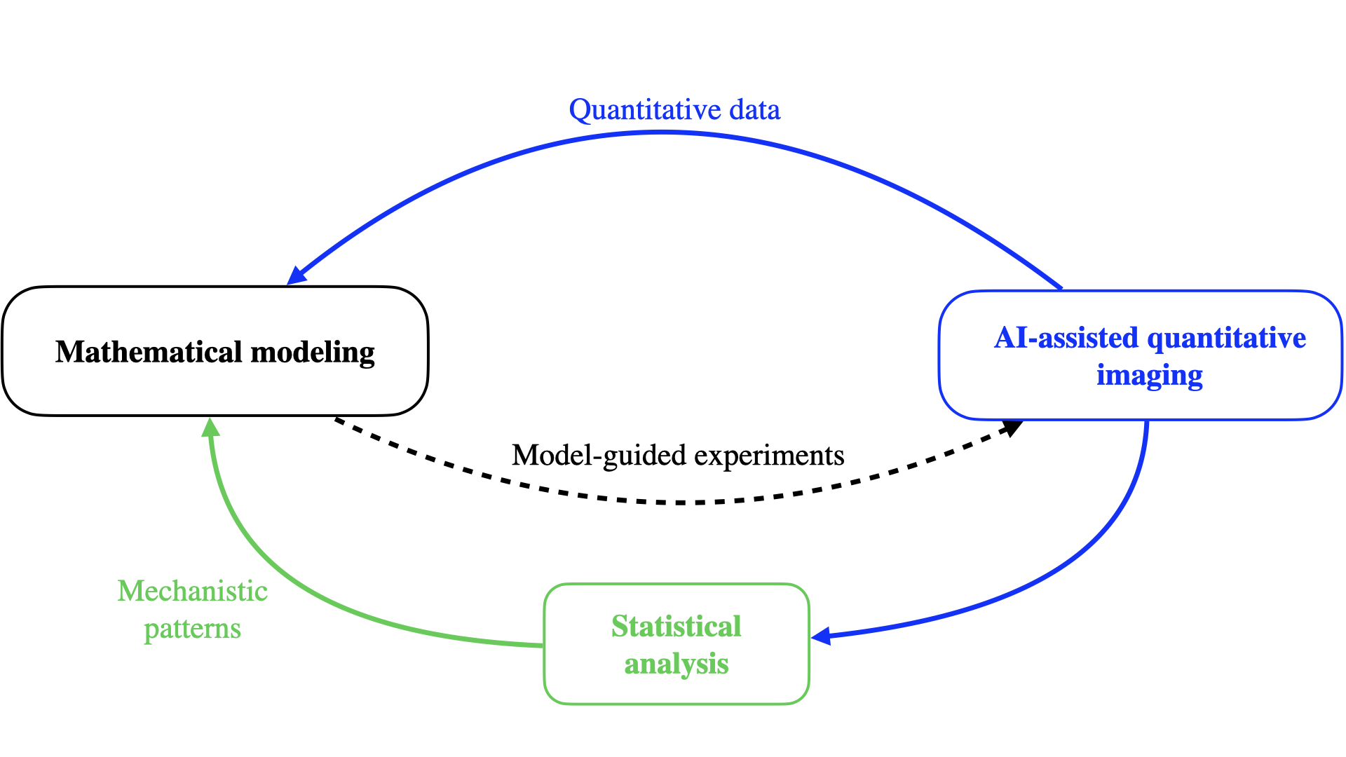

Mathematical modeling of dynamic networks, to explain how stable biological processes emerge from local interactions and to predict how they are perturbed.

Statistical analysis of spatial and temporal relations between agents, to extract mechanistic insights from complex observational datasets.

– AI-assisted image quantification, to transform high-dimensional imaging data into interpretable features that inform models and analyses.


We will apply these approaches to two complementary systems that provide a comparative perspective on biological processes across scales—from single molecules to whole organisms—and how their stability is perturbed by microbial influence.

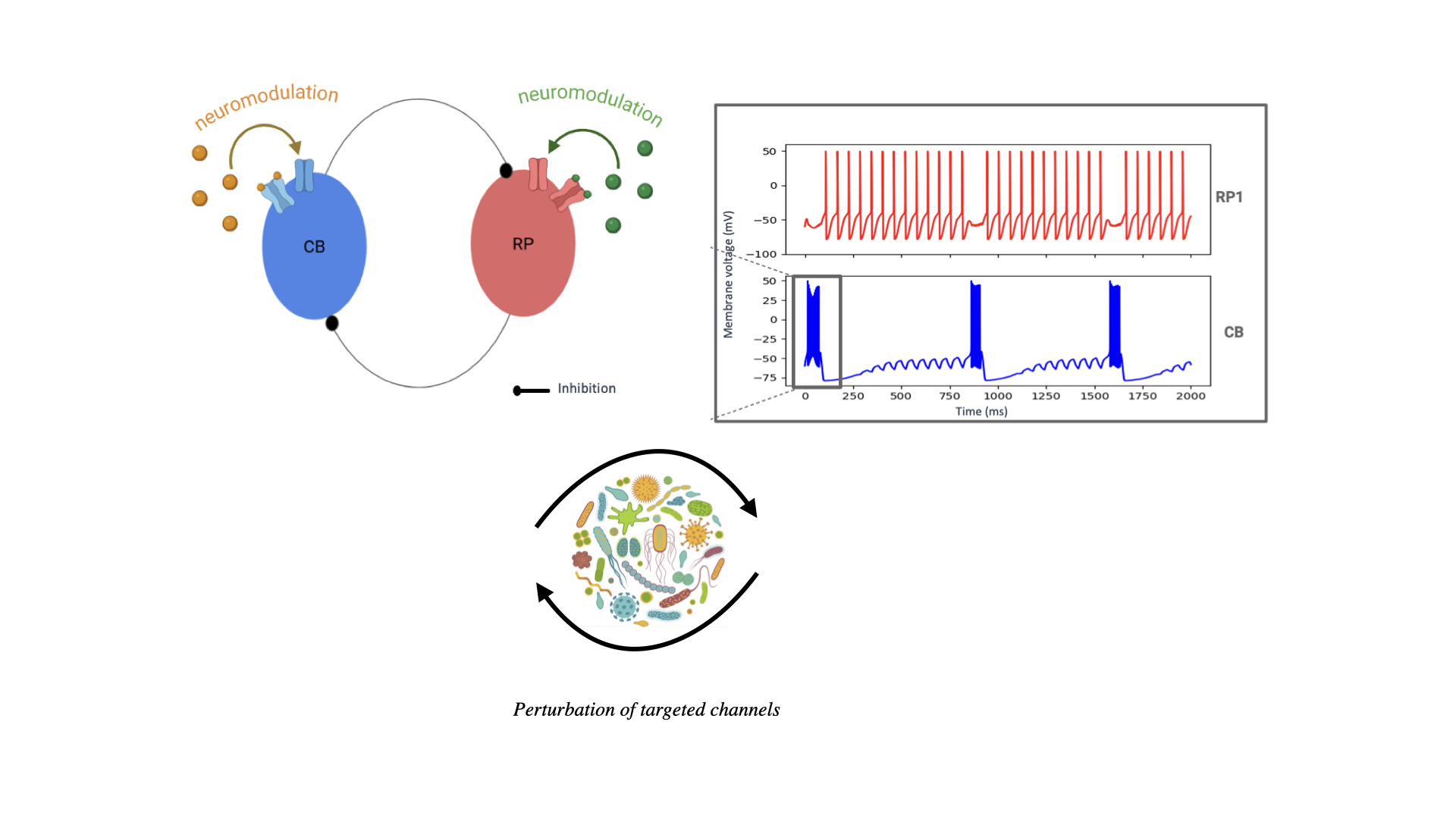
Hydra as a paradigm for neuronal homeostasis.

Calcium imaging of neuronal activity in Hydra

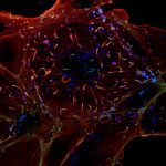
With 200–2,000 neurons spanning 11 cell types, Hydra exhibits diverse quantifiable behaviors such as contractions, locomotion, feeding, and learning. Its rhythmic neural activity makes it a valuable model for studying central pattern generators, which are also relevant to human physiology: recent studies have shown that disruptions of the gut microbiota can impair pacemaker rhythmicity and contribute to abnormal peristalsis. Hydra’s transparency and regenerative capacity make it uniquely suited for long-term imaging and perturbation studies. Using genetically engineered GCaMP Hydra, we aim to monitor neural activity and investigate its modulation by molecular factors such as neuropeptides and the Hydra microbiome.


Host–pathogen interactions at the earliest stages of infection.

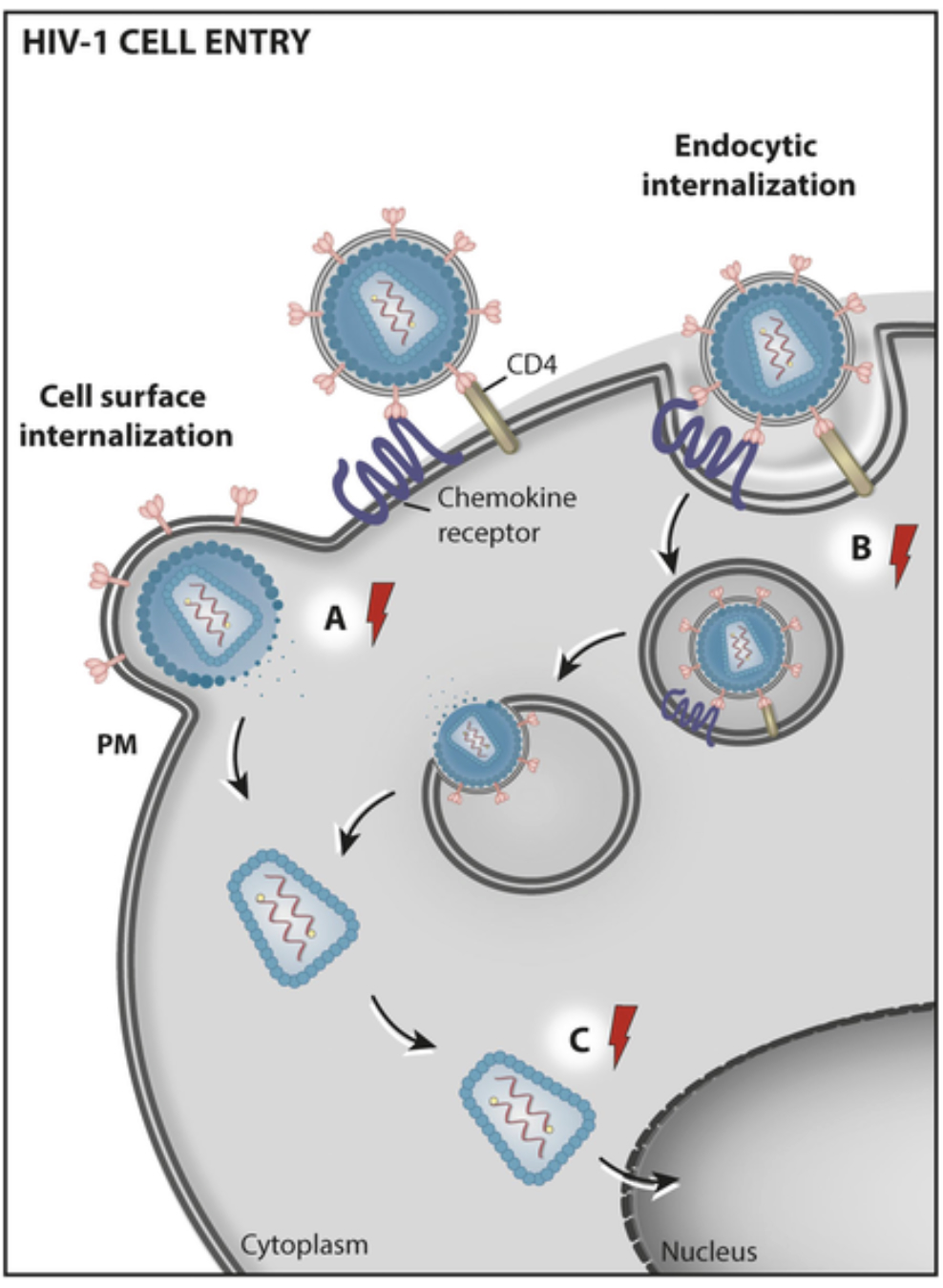
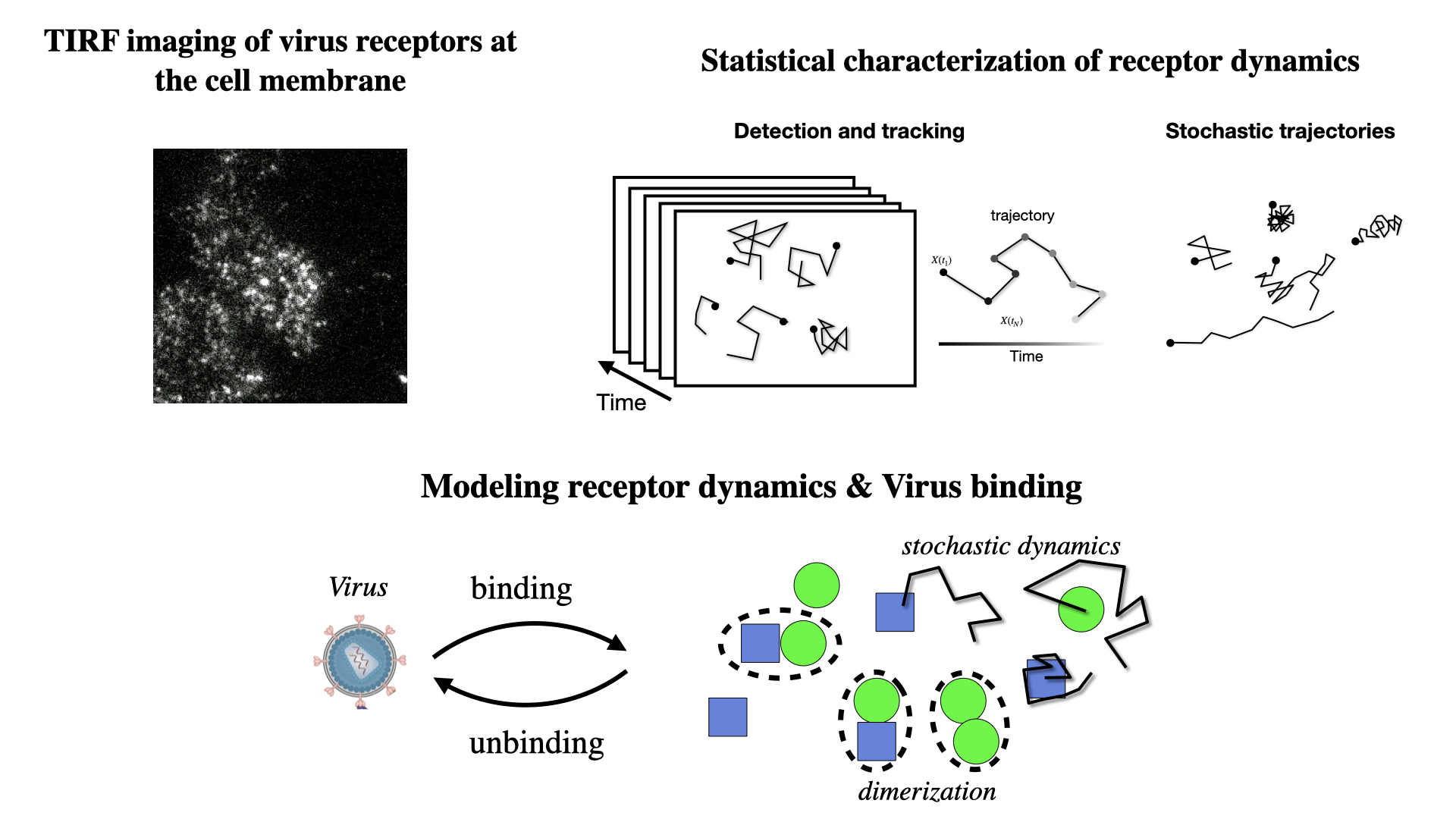
We focus on how viruses such as SARS-CoV-2 and HIV hijack cellular machinery, using advanced live imaging to study receptor and viral dynamics in space and time. These studies are carried out in collaboration with experimental partners specialized in experimental infection biology (e.g. Dynamics of Host–Pathogen Interactions Unit).

HIV hijacking of the cellular trafficking machinery (Adapted from: Tavares, L. et al (2021). Frontiers in Cell and Developmental Biology. 9. 622610. 10.3389/fcell.2021.622610.)

Methodological Axes

1- Mathematical modeling of dynamic networks

Using first-principles and stochastic analyses, we study how stable biological processes emerge from local interactions and how microbes perturb them. In Hydra, we build predictive neuronal models, from minimal oscillators to multi-neuron simulations derived from scRNA-seq data, to analyze how rhythms are modulated by neuropeptides, microbes, and environmental inputs. In parallel, we reconstruct the temporal choreography of viral entry by tracking receptors and viral particles and modeling key biophysical determinants.

2- Statistical analysis of spatial and temporal relations between agents

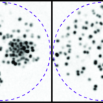
We identify mechanistic patterns in complex datasets by characterizing neuronal ensembles in Hydra and mapping host–pathogen interactions, where receptors, viral particles, and signaling complexes must be coordinated in space and time. Building on our point process framework, we generate multiscale maps linking neuronal firing to behavior and molecular interactions to cellular homeostasis or viral hijacking.

Live imaging and analysis of stochastic receptor dynamics reveal how viruses exploit multiple receptor conformations for cell entry.

3- AI-assisted image quantification

We develop next-generation algorithms to extract quantitative information from imaged processes. In particular, we design tracking algorithms to follow thousands of objects – neurons in Hydra or single virions (or bacteria)  and receptors at membranes. Our framework combine AI-based recognition with probabilistic filtering for robust performance under tissue deformation and crowding.

Former Members

2000
2000
Name
Position
2020
2023
Samuel Kubler
PhD Student, now Postdoc @ Ecole Normale Supérieure, Paris
2018
2021
Suvadip Mukherjee
PostDoc, now Staff scientist @KLA Corporation, Ann Arbor, Michigan, USA

Projects

Spatial Analysis in Biological Imaging

To fully understand protein functions and the intricate molecular networks they form, it’s essential not only to achieve precise detection and tracking of labeled molecules but also to determine their exact localization and spatial proximity. Recent technological advancements in single-molecule labeling and super-resolution multicolor microscopy have revolutionized our ability to observe small molecular clusters that were previously undetectable with conventional microscopy. This capability raises a critical challenge: distinguishing between coincidental proximity—where molecules appear close due to random distribution—and genuine molecular associations. Accurately quantifying these spatial relationships is crucial for identifying meaningful patterns, such as the formation of heterogeneous molecular assemblies. These assemblies are key to understanding how proteins interact in cellular processes, but their detection requires careful analysis to ensure that observed proximity reflects actual functional relationships rather than mere chance.


At the tissue level, recent advancements in digital pathology, such as multi-color immunohistochemistry and multiplex imaging, have significantly enhanced our ability to map the intricate organization of cells within tissues. When coupled with automated image analysis, these techniques offer valuable insights into key areas of research, including cancer biology, immunology, and neuroscience. The integration of spatiotemporal cell mapping enables a more detailed examination of cellular dynamics within complex tissue environments. Understanding the spatial relationships between cells and tissue regions has proven to be a crucial factor in predicting patient outcomes. However, a comprehensive method that effectively links localized cell interactions with the broader tissue context remains an area of ongoing development.

Leveraging level sets to map the spatial relationships between different types of microglial cells in fetal human brain sections.

Main Publications
  • Lagache, T., Grassart, A., Dallongeville, S., Faklaris, O., Sauvonnet, N., Dufour, A., … & Olivo-Marin, J. C. (2018). Mapping molecular assemblies with fluorescence microscopy and object-based spatial statistics. Nature communications9(1), 698.
  • Perochon, T., Krsnik, Z., Massimo, M., Ruchiy, Y.,Lagache ,T*,Manassa D.* & Holcman, D.* (2025). Unraveling microglial spatial organization in the developing human brain with DeepCellMap, a deep learning approach coupled with spatial statistics. Nature Communications16(1), 1577. (* co-corresponding authors)
Links to videos:

A general introduction to Spatial Statistics (Course @ ENS Paris March 2025 :

– Part 1: https://www.youtube.com/watch?v=wVAyrv6uRpM

-Part 2: https://www.youtube.com/watch?v=Xx4Yx_wwNvI

A tutorial for using SODA plugin in Icy for colocalization analysis by Lydia Danglot (IPNP, Paris)

https://www.youtube.com/watch?v=7yVp73s-4TA

Quantitative Neurobiology of Hydra

To understand the origins of disease, we must explore host physiology as a metaorganism—a dynamic system shaped by the interaction and coevolution of host biology, microbes, and the environment. Specifically, my goal is to understand how molecular factors and microbiota influence the homeostasis of neuronal networks and related behaviors in Hydra.

Central to this is decoding the neural code—mapping the neural substrates that drive behavior. In my team, we focus on the small cnidarian Hydra vulgaris, which has a primitive and relatively simple nervous system that could potentially be fully understood. The nervous system consists of 200-2,000 neurons (depending on the size of the animal) that belong to only eleven cell types, organized into two nerve nets without cephalization or ganglia. Hydra exhibits a well-characterized behavioral repertoire, which has been categorized and quantified using machine learning techniques. This repertoire includes simple movements like contractions, twists, and elongations, as well as more complex fixed-action patterns, such as feeding, locomotion through somersaulting and inch-worming, and even some learning paradigms [6]. As a polyp, the Hydra possesses remarkable regenerative capabilities, making it an ideal subject for studying neurodevelopment and the impact of molecular or genetic perturbations on neural connectivity.

From an experimental standpoint, Hydra’s transparency and small size make it an ideal model for microscopy, with nearly all of its neurons suitable for high-speed confocal imaging using calcium indicators. We developed a GCaMP-expressing Hydra colony, and calcium imaging is performed at the institute’s imaging platform.

Although primitive, the molecular toolkit and functional organization of the Hydra nervous system mirror those of more evolved bilaterian organisms. In particular, Hydra’s nerve net activity is organized into coactive neuronal ensembles that can be considered as functional units, similar to the mammalian cerebral cortex. They represent fundamental building blocks, or the “alphabet” of the neural code. The main neuronal ensembles in Hydra are rhythmically activated, providing a useful model for studying central pattern generators and their modulation by neuropeptides and the animal microbiome. This rhythmic activity is particularly relevant in the context of human physiology, as recent studies have shown that disturbances in the gut microbiota can disrupt pacemaker rhythmicity and gut motility, leading to gastrointestinal conditions like irritable bowel syndrome.

To develop a comprehensive framework that would link neuronal activity to behavior, we focus on three main research axis:

  1. Robust, long-term monitoring of single-neuron activity in behaving Hydra
  2. Development of a statistical framework to relate single-neuron activity to the neural substrates underlying behavior.
  3. Creation of an integrated mathematical model and simulation tools that combine imaging and behavioral data to explore how environmental factors (such as light, salinity, etc.) and molecular factors, such as neuropeptides, modulate the functional organization of Hydra’s neural network and behavior.

This approach will not only deepen our understanding of Hydra’s neural function but also contribute to the broader field of integrating and elucidating the influence of various factors on neural development and homeostasis.

Main Publications
  • Hanson, A., Reme, R., Telerman, N., Yamamoto, W., Olivo-Marin, J. C., Lagache, T.*, & Yuste, R.* (2024). Automatic monitoring of neural activity with single-cell resolution in behaving Hydra. Scientific Reports14(1), 5083. (*: co-corresponding authors)
  • Lagache, T., Hanson, A., Pérez-Ortega, J. E., Fairhall, A., & Yuste, R. (2021). Tracking calcium dynamics from individual neurons in behaving animals. PLoS computational biology17(10), e1009432.
Links to videos:

Great video from our collaborator Alison Hanson about the “inner self” of Hydra: https://www.youtube.com/watch?v=bZPhqwrSC24

Transversal Project

Fundings

Publications

Download