Link to Pubmed [PMID] – 32944900
Link to DOI – 10.1007/978-1-0716-0958-3_2
Methods Mol. Biol. 2021 ; 2214(): 11-30
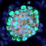
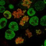
The mouse preimplantation embryo is an excellent system for studying how mammalian cells organize dynamically into increasingly complex structures. Accessible to experimental and genetic manipulations, its normal or perturbed development can be scrutinized ex vivo by real-time imaging from fertilization to late blastocyst stage. High-resolution imaging of multiple embryos at the same time can be compromised by embryos displacement during imaging. We have developed an inexpensive and easy-to-produce imaging device that facilitates greatly the imaging of preimplantation embryo. In this chapter, we describe the different steps of production and storage of the imaging device as well as its use for live imaging of mouse preimplantation embryos expressing fluorescent reporters from genetically modified alleles or after in vitro transcribed mRNA transfer by microinjection or electroporation.


