
![]()
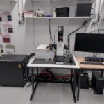
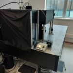
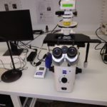
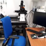
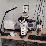
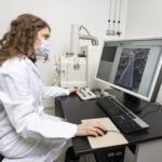
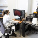
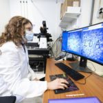
At the Imaging Facility of the Hearing Institute, we offer diverse systems to meet your imaging needs. The available instruments include:
- Epifluorescence microscopes
- Confocal microscopes
- Spinning disk microscopes
- STED microscopes
- Two-photon microscopes
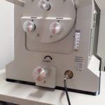
- Benchtop electron microscope
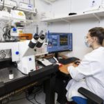
These instruments allow you to observe samples at various levels, ranging from entire organoids to the fine details of intracellular structures (~50 nm lateral resolution in optical microscopy).
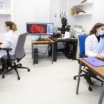
We provide support and services in the image acquisition process for both fixed and live samples. We also assist with image treatment, analysis, and interpretation.
We strive to offer guidance in sample preparation and manage the cell culture and histology rooms. Our goal is to establish protocols for sample preparation and labeling tailored to the typical research activities of the Hearing Institute, also work with the institute’s teams to develop home-made optical systems tailored for live imaging. We offer these resources to both the facility and individual research teams.