


About
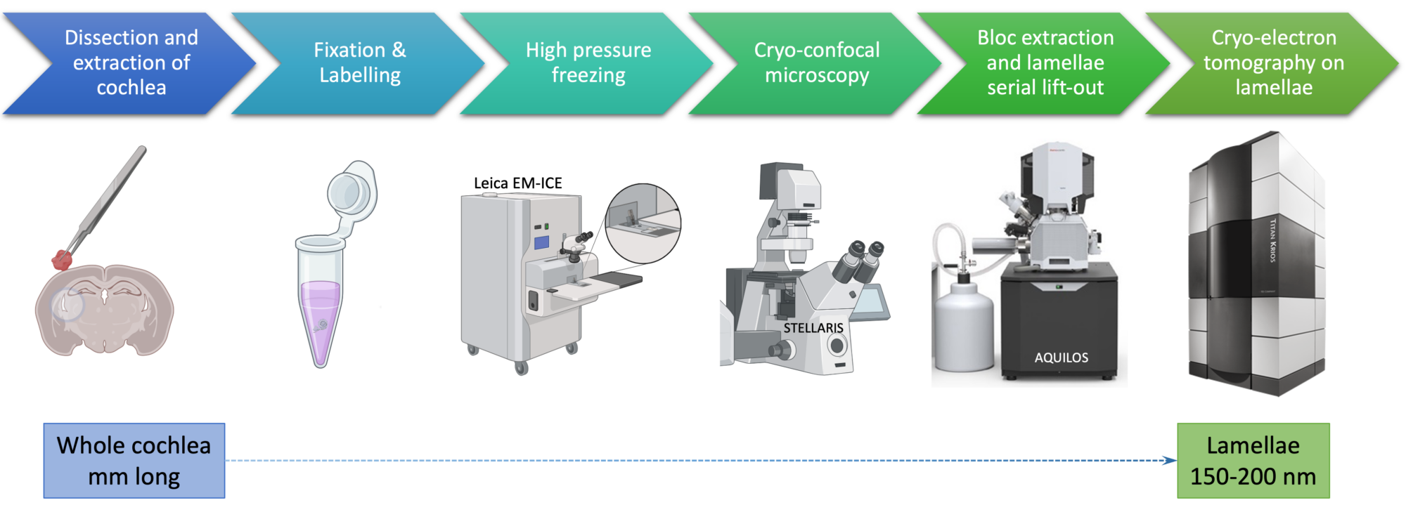
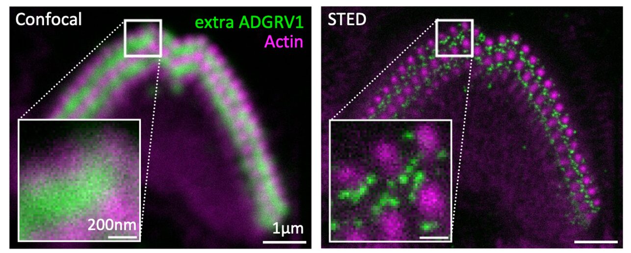
The project aims to address the general structure in situ of the native protein complex Usher 2, needed for the development of the cochlea, the organ of hearing, and to elucidate the impact of deafness mutations in pathological processes. The Usher 2 syndrome is the most common form of hereditary hearing-vision loss in humans. We propose to combine cutting-edge imaging approaches using cryo-electron tomography (cryoET), cryo-soft-X-ray tomography (cryoSXT) and super-resolution fluorescence microscopy (STED) on mouse cochlea samples to dissect the 3D organisation of the Usher 2 complex in its native tissue environment. Combined with the atomic resolution structures already obtained in the lab by integrative approaches (Cryo-EM, NMR and X-ray diffraction), we will then be able to provide a comprehensive view of the Usher 2 complex in situ.
Altogether, our structural results from the micro-microscale (cochlea) to the nano- and atomic scale (protein domains) will lead us to propose a model on how Usher 2 proteins and partners get organised in situ to form a protein network essential for the correct cochlea development. This molecular characterisation will have a potential impact on Usher 2 syndrome diagnosis and provide a deeper understanding of its physiopathology.