About
Congenital heart surgery is a challenging surgical disciplines due to the broad spectrum of conditions and high variability of patient anatomies. Extensive understanding of the complex spatial relationship between anatomical structures is mandatory to plan the optimal surgical approach and therefore reduce operative time, morbidity, and mortality. The last 5 years, we have seen significant accelerations in advancements in the field of innovative three‐dimensional (3D) visualization techniques due to the ever‐growing availability of 3D‐ready imaging data derived from cardiac nuclear magnetic resonance, cardiac computed tomography (CT) scan, and 3D echocardiography.
However, the spread of these technologies has been limited by the lack of standardized approaches, long processing times, high costs, and a lack of dynamic representations of the cardiac cycle without any hemodynamic data. More importantly, there are strong limitations and heterogeneities in teaching congenital heart disease diagnosis and surgeries in Europe (& all around the world). Improving patient care requires introduction of these technologies at the earliest stage of medical student training.
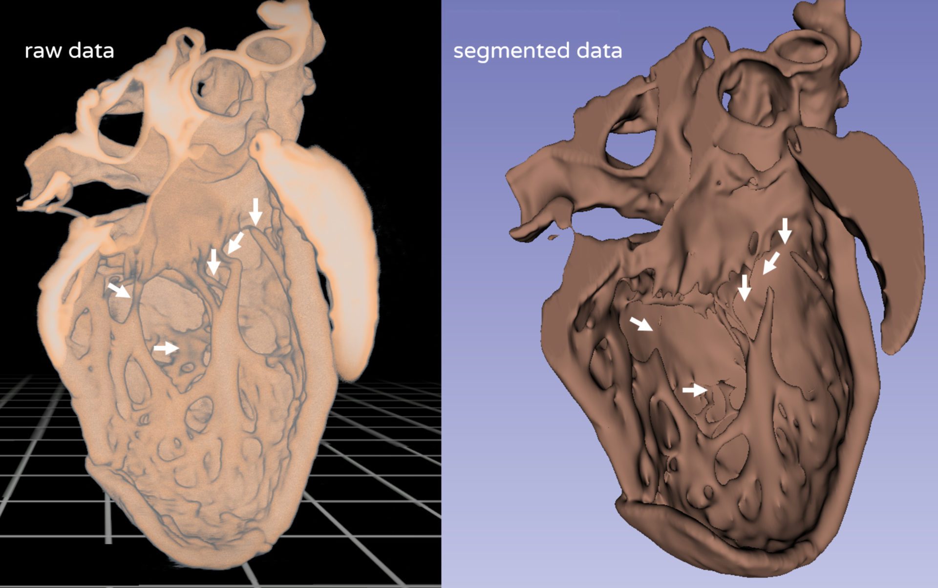
Example of Comparison of a full volumetric rendering of a heart CT-scan vs segmented heart.
Example of full volumetric representation of a heart from high intensity CT-scan imaging without segmentation.


