Search anything and hit enter
  • Teams
  • Members
  • Projects
  • Events
  • Calls
  • Jobs
  • publications
  • Software
  • Tools
  • Network
  • Equipment

A little guide for advanced search:

  • Tip 1. You can use quotes "" to search for an exact expression.
    Example: "cell division"
  • Tip 2. You can use + symbol to restrict results containing all words.
    Example: +cell +stem
  • Tip 3. You can use + and - symbols to force inclusion or exclusion of specific words.
    Example: +cell -stem
e.g. searching for members in projects tagged cancer
Search for
Count
IN
OUT
Content 1
  • member
  • team
  • department
  • center
  • program_project
  • nrc
  • whocc
  • project
  • software
  • tool
  • patent
  • Administrative Staff
  • Assistant Professor
  • Associate Professor
  • Clinical Research Assistant
  • Clinical Research Nurse
  • Clinician Researcher
  • Department Manager
  • Dual-education Student
  • Full Professor
  • Honorary Professor
  • Lab assistant
  • Master Student
  • Non-permanent Researcher
  • Nursing Staff
  • Permanent Researcher
  • Pharmacist
  • PhD Student
  • Physician
  • Post-doc
  • Prize
  • Project Manager
  • Research Associate
  • Research Engineer
  • Retired scientist
  • Technician
  • Undergraduate Student
  • Veterinary
  • Visiting Scientist
  • Deputy Director of Center
  • Deputy Director of Department
  • Deputy Director of National Reference Center
  • Deputy Head of Facility
  • Director of Center
  • Director of Department
  • Director of Institute
  • Director of National Reference Center
  • Group Leader
  • Head of Facility
  • Head of Operations
  • Head of Structure
  • Honorary President of the Departement
  • Labex Coordinator
Content 2
  • member
  • team
  • department
  • center
  • program_project
  • nrc
  • whocc
  • project
  • software
  • tool
  • patent
  • Administrative Staff
  • Assistant Professor
  • Associate Professor
  • Clinical Research Assistant
  • Clinical Research Nurse
  • Clinician Researcher
  • Department Manager
  • Dual-education Student
  • Full Professor
  • Honorary Professor
  • Lab assistant
  • Master Student
  • Non-permanent Researcher
  • Nursing Staff
  • Permanent Researcher
  • Pharmacist
  • PhD Student
  • Physician
  • Post-doc
  • Prize
  • Project Manager
  • Research Associate
  • Research Engineer
  • Retired scientist
  • Technician
  • Undergraduate Student
  • Veterinary
  • Visiting Scientist
  • Deputy Director of Center
  • Deputy Director of Department
  • Deputy Director of National Reference Center
  • Deputy Head of Facility
  • Director of Center
  • Director of Department
  • Director of Institute
  • Director of National Reference Center
  • Group Leader
  • Head of Facility
  • Head of Operations
  • Head of Structure
  • Honorary President of the Departement
  • Labex Coordinator
Search

← Go to Research

Go back
Scroll to top
Share
Scientific Fields
Diseases
Organisms
Applications
Technique
Starting Date
01
Feb 2015
Ending Date
01
Dec 2020
Status
Completed
Members
4
Structures
2
Publications
6

About

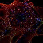
Elucidating the mechanisms underlying cell deformation and motility is a topic of major interest in cell biology, for they are strongly involved in cell development, immune responses, cancer and infectious diseases. This projects focuses on the development robust and fully automated image analysis tools, able to extract quantitative measures of cell architecture, shape and motility from multi-dimensional (2D/3D), multi-modal (brightfield/phase-contrast/fluorescence) time-lapse microscopy data, with the aim to derive mathematical models of 3D cellular morphodynamics.

Working model: Entamoeba histolytica

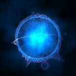
Entamoeba histolytica, the causative agent of human amoebiasis, is a protozoan parasite characterised by its amoeboid motility, which is essential to its survival and invasion of the human host. Elucidating the molecular mechanisms leading to invasion of human tissues by of E. histolytica requires a quantitative understanding of how its cytoskeleton deforms and tailors its mode of migration to the local microenvironment. In this project we develop a fully interdisciplinary setting to tackle this challenge, placed at the interface between cell biology, biochemistry, fluorescence probe development, live cell imaging, and computational analysis & modelling, with the ultimate goal to decipher the morphodynamics and biomechanics of amoeboid motion in multiple experimental contexts, from simple in vitro models to complex reconstituted multi-cellular matrices.
Material and methods

In order to precisely segment and extract the cell shape from image data, we have been focusing on the past years on the theory of deformable models, also known as “active contours” techniques. The principle is to deform an initial contour placed on the image until it fits the boundary of the target cell. The deformation can be mathematically expressed as the minimization of an energy functional, which comprises several terms related either to the image data (driving the contour toward the cell boundary) or to geometrical properties of the contour (regularizing the deformation to avoid local energy minima). This energy is an essential ingredient of the method, and several of our contributions consist in adapting this energy and its implementation to meet the particular constraints of biological imaging, e.g., low signal-to-noise ratio, multiple cell-cell contacts over time, inhomogeneous cell staining, photo-bleaching, etc.. Active contours also offer a flexible formalism for efficient shape representation and quantification, allowing to compute robust statistics over large-scale cell populations. The algorithms developed for this project are currently used through collaborations for numerous applications including multi-cell segmentation, tracking, interaction and particle localization.

Fundings

References