Lien vers Pubmed [PMID] – 26484791
Lien DOI – 10.3791/53197
J Vis Exp 2015 Oct; (104):
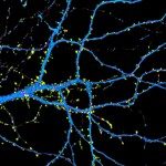
Dendritic spines are small protrusions that correspond to the post-synaptic compartments of excitatory synapses in the central nervous system. They are distributed along the dendrites. Their morphology is largely dependent on neuronal activity, and they are dynamic. Dendritic spines express glutamatergic receptors (AMPA and NMDA receptors) on their surface and at the levels of postsynaptic densities. Each spine allows the neuron to control its state and local activity independently. Spine morphologies have been extensively studied in glutamatergic pyramidal cells of the brain cortex, using both in vivo approaches and neuronal cultures obtained from rodent tissues. Neuropathological conditions can be associated to altered spine induction and maturation, as shown in rodent cultured neurons and one-dimensional quantitative analysis (1). The present study describes a protocol for the 3D quantitative analysis of spine morphologies using human cortical neurons derived from neural stem cells (late cortical progenitors). These cells were initially obtained from induced pluripotent stem cells. This protocol allows the analysis of spine morphologies at different culture periods, and with possible comparison between induced pluripotent stem cells obtained from control individuals with those obtained from patients with psychiatric diseases.


