Présentation
Adaptive immune responses are initiated in secondary lymphoid organs through cellular interactions between antigen-specific T cells and antigen-presenting dendritic cells. One of the aim of our lab is to better understand how T cells collect activation signals in vivo. Using intravital multiphoton imaging, we are therefore studying how T cell-DC contacts are regulated and how activation signals are delivered during individual interactions. These studies help identify fundamental mechanisms shaping the efficacy of T cell activation and differentiation. Recently, we have extended these questions to natural killer cells.

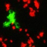
T cells forming stables interactions with dendritic cells in the lymph node upon antigen recognition. T cells (red) and dendritic cells (green) are imaged in the popliteal lymph node of a live mouse. Antigen is delivered to DCs at t=0.
Click here to see the movie

Dynamics of the immunological synapse during T cell-B cell interactions in the lymph node. T cells expressed a LAT-GFP fusion protein. B cells are shown in red.
Click here to see the movie
 T cell functional heterogeneity is initiated prior to the first cell division. T cells isolated from an IFN-g YFP reporter mice and labeled with ta red are imaged during their first division after activation. Yellow and green T cells have activated the IFN-g gene at the time of division, whereas red T cells have not.
Click here to see the movie

In vivo imaging TCR signaling in real-time using CD62L shedding. GFP-expressing T cells (green) are labeled in vivo with fluorescent anti-CD62L Fab (red). Antigen is delivered at t=0 resulting in T cell stop and progressive decrease of CD62L staining. Intravital imaging was performed in the spleen.
Click here to see the movie
 NK cell motility in lymph nodes. NK cells (red), T cells (blue) and DCs (green) are imaged at steady state in the lymph node.
Click here to see the movie


