Tapez votre recherche ici
  • Équipes
  • Membres
  • Projets
  • Événements
  • Appels
  • Emplois
  • publications
  • Logiciel
  • Outils
  • Réseau
  • Équipement

Un petit guide pour l'utilisation de la recherche avancée :

  • Tip 1. Utilisez "" afin de chercher une expression exacte.
    Exemple : "division cellulaire"
  • Tip 2. Utilisez + afin de rendre obligatoire la présence d'un mot.
    Exemple : +cellule +stem
  • Tip 3. Utilisez + et - afin de forcer une inclusion ou exclusion d'un mot.
    Exemple : +cellule -stem
e.g. searching for members in projects tagged cancer
Rechercher
Compteur
IN
OUT
Contenu 1
  • member
  • team
  • department
  • center
  • program_project
  • nrc
  • whocc
  • project
  • software
  • tool
  • patent
  • Personnel Administratif
  • Chargé(e) de Recherche Expert
  • Directeur(trice) de Recherche
  • Assistant(e) de Recherche Clinique
  • Infirmier(e) de Recherche Clinique
  • Chercheur(euse) Clinicien(ne)
  • Manager de département
  • Etudiant(e) en alternance
  • Professeur(e)
  • Professeur Honoraire
  • Aide technique
  • Etudiant(e) M2
  • Chercheur(euse) Contractuel(le)
  • Personnel infirmier
  • Chercheur(euse) Permanent(e)
  • Pharmacien(ne)
  • Etudiant(e) en thèse
  • Médecin
  • Post-doctorant(e)
  • Prize
  • Chef(fe) de Projet
  • Chargé(e) de Recherche
  • Ingénieur(e) de Recherche
  • Chercheur(euse) Retraité(e)
  • Technicien(ne)
  • Etudiant(e)
  • Vétérinaire
  • Visiteur(euse) Scientifique
  • Directeur(trice) Adjoint(e) de Centre
  • Directeur(trice) Adjoint(e) de Départment
  • Directeur(trice) Adjoint(e) de Centre National de Référence
  • Directeur(trice) Adjoint(e) de Plateforme
  • Directeur(trice) de Centre
  • Directeur(trice) de Départment
  • Directeur(trice) d'Institut
  • Directeur(trice) de Centre National de Référence
  • Chef(fe) de Groupe
  • Responsable de Plateforme
  • Responsable opérationnel et administratif
  • Responsable de Structure
  • Président(e) d'honneur de Département
  • Coordinateur(trice) du Labex
Contenu 2
  • member
  • team
  • department
  • center
  • program_project
  • nrc
  • whocc
  • project
  • software
  • tool
  • patent
  • Personnel Administratif
  • Chargé(e) de Recherche Expert
  • Directeur(trice) de Recherche
  • Assistant(e) de Recherche Clinique
  • Infirmier(e) de Recherche Clinique
  • Chercheur(euse) Clinicien(ne)
  • Manager de département
  • Etudiant(e) en alternance
  • Professeur(e)
  • Professeur Honoraire
  • Aide technique
  • Etudiant(e) M2
  • Chercheur(euse) Contractuel(le)
  • Personnel infirmier
  • Chercheur(euse) Permanent(e)
  • Pharmacien(ne)
  • Etudiant(e) en thèse
  • Médecin
  • Post-doctorant(e)
  • Prize
  • Chef(fe) de Projet
  • Chargé(e) de Recherche
  • Ingénieur(e) de Recherche
  • Chercheur(euse) Retraité(e)
  • Technicien(ne)
  • Etudiant(e)
  • Vétérinaire
  • Visiteur(euse) Scientifique
  • Directeur(trice) Adjoint(e) de Centre
  • Directeur(trice) Adjoint(e) de Départment
  • Directeur(trice) Adjoint(e) de Centre National de Référence
  • Directeur(trice) Adjoint(e) de Plateforme
  • Directeur(trice) de Centre
  • Directeur(trice) de Départment
  • Directeur(trice) d'Institut
  • Directeur(trice) de Centre National de Référence
  • Chef(fe) de Groupe
  • Responsable de Plateforme
  • Responsable opérationnel et administratif
  • Responsable de Structure
  • Président(e) d'honneur de Département
  • Coordinateur(trice) du Labex
Recherche

← Go to Research

Revenir
Haut de page
Partagez
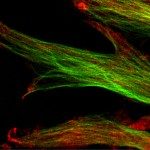
© Sandrine Etienne-Manneville
Photo prise à l'avant (dans la protrusion) d'astrocytes primaires de rat en migration. Marquage par immunofluorescence montrant en rouge, p150 Glued, une protéine associée aux extrémités 'plus' des microtubules et en vert la tubuline des microtubules. La photographie montre l'accumulation de p150 Glued à l'avant des cellules en migration, où la protéine pourrait participer à l'ancrage des microtubules à la membrane plasmique. Pour essayer de corriger, les dérèglements observés lors de la migration des cellules d'astrocytes tumuraux ou gliomes on cherche à connaitre les mécanismes moléculaires fondamentaux qui controlent la polarisation et la migration cellulaires.
Publication : Methods in molecular biology (Clifton, N.J.)

Defining Membrane Protein Topology Using pho-lac Reporter Fusions

Domaines Scientifiques
Maladies
Organismes
Applications
Technique

Publié sur Methods in molecular biology (Clifton, N.J.) - 01 janv. 2017

Karimova G, Ladant D

Lien vers Pubmed [PMID] – 28667608

Methods Mol. Biol. 2017;1615:129-142

Experimental determination of membrane protein topology can be achieved using various techniques. Here we present the pho-lac dual reporter system, a simple, convenient, and reliable tool to analyze the topology of membrane proteins in vivo. The system is based on the use of two topological markers with complementary properties, the Escherichia coli β-galactosidase LacZ, which is active in the cytoplasm, and the E. coli alkaline phosphatase PhoA, which is active in the bacterial periplasm. Specifically, in this pho-lac gene system, the reporter molecule is a chimera composed of the mature PhoA that is in frame with the β-galactosidase α-peptide, LacZα. Hence, when targeted to the periplasm, the PhoA-LacZα dual reporter displays high alkaline phosphatase activity but no β-galactosidase activity. Conversely, when located in the cytoplasm, PhoA-LacZα has no phosphatase activity but exhibits high β-galactosidase activity in E. coli cells expressing the ω fragment of LacZ, LacZω (via the α-complementation phenomenon). The dual nature of the PhoA-LacZα reporter allows a simple way to normalize both enzymatic activities to obtain readily interpretable information about the subcellular location of the fusion site between the membrane protein under study and the reporter. In addition, the PhoA-LacZα reporter permits utilization of dual-indicator agar plates to easily discriminate between colonies bearing cytoplasmic fusions, periplasmic fusions, or out-of-frame fusions. In total, the phoA-lacZα fusion reporter approach is a straightforward and rather inexpensive method of characterizing the topology of membrane proteins in vivo.