Lien vers Pubmed [PMID] – 22683534
Toxicon 2012 Sep;60(4):531-8
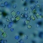
This review will focus on a description of the three-dimensional structures of two β-neurotoxins, the monomeric PLA(2) ammodytoxin from Vipera ammodytes ammodytes, and heterodimeric crotoxin from Crotalus durissus terrificus, and a detailed structural analysis of their multiple functional sites. We have recently determined at high resolution the crystal structures of two natural isoforms of ammodytoxin (AtxA and AtxC) (Saul et al., 2010) which exhibit different toxicity profiles and different anticoagulant properties. Comparative structural analysis of these two PLA(2) isoforms, which differ only by two amino acid residues, allowed us to detect local conformational changes and delineate the role of critical residues in the anticoagulant and neurotoxic functions of these PLA(2) (Saul et al., 2010). We have also determined, at 1.35Å resolution, the crystal structure of heterodimeric crotoxin (Faure et al., 2011). The three-dimensional structure of crotoxin revealed details of the binding interface between its acidic (CA) and basic (CB) subunits and allowed us to identify key residues involved in the stability and toxicity of this potent heterodimeric β-neurotoxin (Faure et al., 2011). The precise spatial orientation of the three covalently linked polypeptide chains in the mature CA subunit complexed with CB helps us to understand the role played by critical residues of the CA subunit in the increased toxicity of the crotoxin complex. Since the CA subunit is a natural inhibitor of the catalytic and anticoagulant activities of CB, identification of the CA-CB binding interface describes residues involved in this inhibition. We propose future research directions based on knowledge of the recently reported 3D structures of crotoxin and ammodytoxin.

