Search anything and hit enter
  • Teams
  • Members
  • Projects
  • Events
  • Calls
  • Jobs
  • publications
  • Software
  • Tools
  • Network
  • Equipment

A little guide for advanced search:

  • Tip 1. You can use quotes "" to search for an exact expression.
    Example: "cell division"
  • Tip 2. You can use + symbol to restrict results containing all words.
    Example: +cell +stem
  • Tip 3. You can use + and - symbols to force inclusion or exclusion of specific words.
    Example: +cell -stem
e.g. searching for members in projects tagged cancer
Search for
Count
IN
OUT
Content 1
  • member
  • team
  • department
  • center
  • program_project
  • nrc
  • whocc
  • project
  • software
  • tool
  • patent
  • Administrative Staff
  • Assistant Professor
  • Associate Professor
  • Clinical Research Assistant
  • Clinical Research Nurse
  • Clinician Researcher
  • Department Manager
  • Dual-education Student
  • Full Professor
  • Honorary Professor
  • Lab assistant
  • Master Student
  • Non-permanent Researcher
  • Nursing Staff
  • Permanent Researcher
  • Pharmacist
  • PhD Student
  • Physician
  • Post-doc
  • Prize
  • Project Manager
  • Research Associate
  • Research Engineer
  • Retired scientist
  • Technician
  • Undergraduate Student
  • Veterinary
  • Visiting Scientist
  • Deputy Director of Center
  • Deputy Director of Department
  • Deputy Director of National Reference Center
  • Deputy Head of Facility
  • Director of Center
  • Director of Department
  • Director of Institute
  • Director of National Reference Center
  • Group Leader
  • Head of Facility
  • Head of Operations
  • Head of Structure
  • Honorary President of the Departement
  • Labex Coordinator
Content 2
  • member
  • team
  • department
  • center
  • program_project
  • nrc
  • whocc
  • project
  • software
  • tool
  • patent
  • Administrative Staff
  • Assistant Professor
  • Associate Professor
  • Clinical Research Assistant
  • Clinical Research Nurse
  • Clinician Researcher
  • Department Manager
  • Dual-education Student
  • Full Professor
  • Honorary Professor
  • Lab assistant
  • Master Student
  • Non-permanent Researcher
  • Nursing Staff
  • Permanent Researcher
  • Pharmacist
  • PhD Student
  • Physician
  • Post-doc
  • Prize
  • Project Manager
  • Research Associate
  • Research Engineer
  • Retired scientist
  • Technician
  • Undergraduate Student
  • Veterinary
  • Visiting Scientist
  • Deputy Director of Center
  • Deputy Director of Department
  • Deputy Director of National Reference Center
  • Deputy Head of Facility
  • Director of Center
  • Director of Department
  • Director of Institute
  • Director of National Reference Center
  • Group Leader
  • Head of Facility
  • Head of Operations
  • Head of Structure
  • Honorary President of the Departement
  • Labex Coordinator
Search
Go back
Scroll to top
Share
© Aline Bonnet, Institut Pasteur
Coupe transversale d’embryon de caille transgénique mbGFP à 18somites, au niveau du futur bourgeon de membre antérieur avec un marquage noyaux (bleu), GFP (vert) et actine (rouge) / Transversal section of a mbGFP transgenic quail embryo at 18-somite stage, at forelimb level, with nuclei (blue), GFP (green) and actin (red) labelling
Publication : Developmental cell

A two-step mechanism for myotome formation in chick

Scientific Fields
Diseases
Organisms
Applications
Technique

Published in Developmental cell - 01 Jun 2004

Gros J, Scaal M, Marcelle C

Link to Pubmed [PMID] – 15177035

Dev. Cell 2004 Jun;6(6):875-82

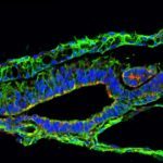
The study of the morphogenetic cell movements underlying myotome formation in the chick embryo has led to the emergence of highly controversial models. Here we report a real-time cell lineage analysis of myotome development using electroporation of a GFP reporter in newly formed chick somites. Confocal analysis of cell movements demonstrates that myotome formation involves two sequential steps. In a first phase, incremental myotome growth results from a contribution of myocytes derived solely from the medial border of the dermomyotome. In a second phase, myocytes are produced from all four borders of the dermomyotome. The relative distribution of myocytes demonstrates that the medial and the lateral borders of the somite generate exclusively epaxial and hypaxial muscles. This analysis also identified five myotomal regions, characterized by the origin of the myocytes that constitute them. Together, our results provide a comprehensive model describing the morphogenesis of the early myotome in higher vertebrates.