Search anything and hit enter
  • Teams
  • Members
  • Projects
  • Events
  • Calls
  • Jobs
  • publications
  • Software
  • Tools
  • Network
  • Equipment

A little guide for advanced search:

  • Tip 1. You can use quotes "" to search for an exact expression.
    Example: "cell division"
  • Tip 2. You can use + symbol to restrict results containing all words.
    Example: +cell +stem
  • Tip 3. You can use + and - symbols to force inclusion or exclusion of specific words.
    Example: +cell -stem
e.g. searching for members in projects tagged cancer
Search for
Count
IN
OUT
Content 1
  • member
  • team
  • department
  • center
  • program_project
  • nrc
  • whocc
  • project
  • software
  • tool
  • patent
  • Administrative Staff
  • Assistant Professor
  • Associate Professor
  • Clinical Research Assistant
  • Clinical Research Nurse
  • Clinician Researcher
  • Department Manager
  • Dual-education Student
  • Full Professor
  • Honorary Professor
  • Lab assistant
  • Master Student
  • Non-permanent Researcher
  • Nursing Staff
  • Permanent Researcher
  • Pharmacist
  • PhD Student
  • Physician
  • Post-doc
  • Prize
  • Project Manager
  • Research Associate
  • Research Engineer
  • Retired scientist
  • Technician
  • Undergraduate Student
  • Veterinary
  • Visiting Scientist
  • Deputy Director of Center
  • Deputy Director of Department
  • Deputy Director of National Reference Center
  • Deputy Head of Facility
  • Director of Center
  • Director of Department
  • Director of Institute
  • Director of National Reference Center
  • Group Leader
  • Head of Facility
  • Head of Operations
  • Head of Structure
  • Honorary President of the Departement
  • Labex Coordinator
Content 2
  • member
  • team
  • department
  • center
  • program_project
  • nrc
  • whocc
  • project
  • software
  • tool
  • patent
  • Administrative Staff
  • Assistant Professor
  • Associate Professor
  • Clinical Research Assistant
  • Clinical Research Nurse
  • Clinician Researcher
  • Department Manager
  • Dual-education Student
  • Full Professor
  • Honorary Professor
  • Lab assistant
  • Master Student
  • Non-permanent Researcher
  • Nursing Staff
  • Permanent Researcher
  • Pharmacist
  • PhD Student
  • Physician
  • Post-doc
  • Prize
  • Project Manager
  • Research Associate
  • Research Engineer
  • Retired scientist
  • Technician
  • Undergraduate Student
  • Veterinary
  • Visiting Scientist
  • Deputy Director of Center
  • Deputy Director of Department
  • Deputy Director of National Reference Center
  • Deputy Head of Facility
  • Director of Center
  • Director of Department
  • Director of Institute
  • Director of National Reference Center
  • Group Leader
  • Head of Facility
  • Head of Operations
  • Head of Structure
  • Honorary President of the Departement
  • Labex Coordinator
Search
Go back
Scroll to top
Share
© Research
Project

Electron Tomography

Scientific Fields
Diseases
Organisms
Applications
Technique
Starting Date
10
Feb 2015
Status
Ongoing
Structures
1

About

Electron tomography (ET) is one of the two EM methods to obtain three- dimensional ultra-structural images. It relies on tilting the specimen and the automated acquisition of multiple tilt projections. It overcomes one of the major limitations of thin sections TEM that is restricted to 2D-images.

proj4
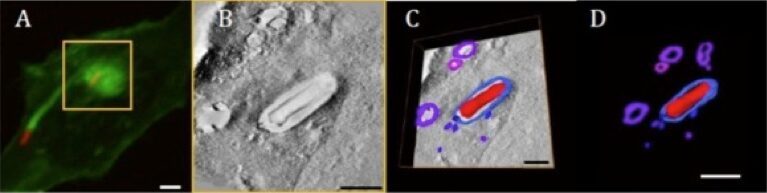
Study of Shigella flexneri infection of Hela cells by 3D Correlative Light-Electron Tomography: in order to identify and image the subsequent entry steps of Shigella into host cells, we developed an innovative methodology that combines correlative light and transmission electron microscopy with serial sectioning and tomography on identical samples. The final aim is to couple the functional information derived by 3D confocal microscopy with the high-resolution structural information from 3D electron tomography. Bacteria entry events identified by actin foci were imaged in 3D by confocal microscopy (fig.A, boxed area: bacteria are labelled in red and GFP-actin in green), processed for transmission electron microscopy and the same entry event were then imaged in 3D by electron tomography (fig.B). The reconstructed tomographic volume was then modelled in 3D (fig.C & D), highlighting novel vesicular structures that are docking or fusing with the phagocytic vacuole. (Scale bars: 1 µm)