Lien vers Pubmed [PMID] – 15890277
J. Struct. Biol. 2005 Jun;150(3):284-99
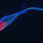
A structural model of the murine PrP small beta-sheet was obtained by synthesizing the RGYMLGSADPNGNQVYYRG peptide comprising the two beta-strands 127-133 and 159-164 linked by a four-residue sequence of high turn propensity. The DPNG turn sequence is a “short circuit” replacing the original protein sequence between the two strands. This 19-residue peptide spontaneously forms very long single fibrils as observed by electron microscopy. The X-ray diffraction patterns of a partially oriented sample reveals an average arrangement of the hairpin peptides into a structure which can be geometrically approximated by an empty-core cylinder. The hairpins are oriented perpendicular to the cylinder axis and a 130 A helix period is observed. Based on X-ray diffraction constraints and on more indirect general protein structure considerations, a precise and consistent fibril model was built. The structure consists of two beta-sheet ribbons wound around a cylinder and assembled into a single fibril with a hairpin orientation perpendicular to the fibril axis. Subsequent implicit and explicit solvent molecular dynamics simulations provided the final structure at atomic resolution and further insights into the stabilizing interactions. Particularly important are the zipper-like network of polar interactions between the edges of the two ribbons, including the partially buried water molecules. The hydrophobic core is not optimally compact explaining the low density of this region seen by X-ray diffraction. The present findings provide also a simple model for further investigating the sequence-stability relationship using a mutational approach with a quasi-independent consideration of the polar and apolar interactions.

