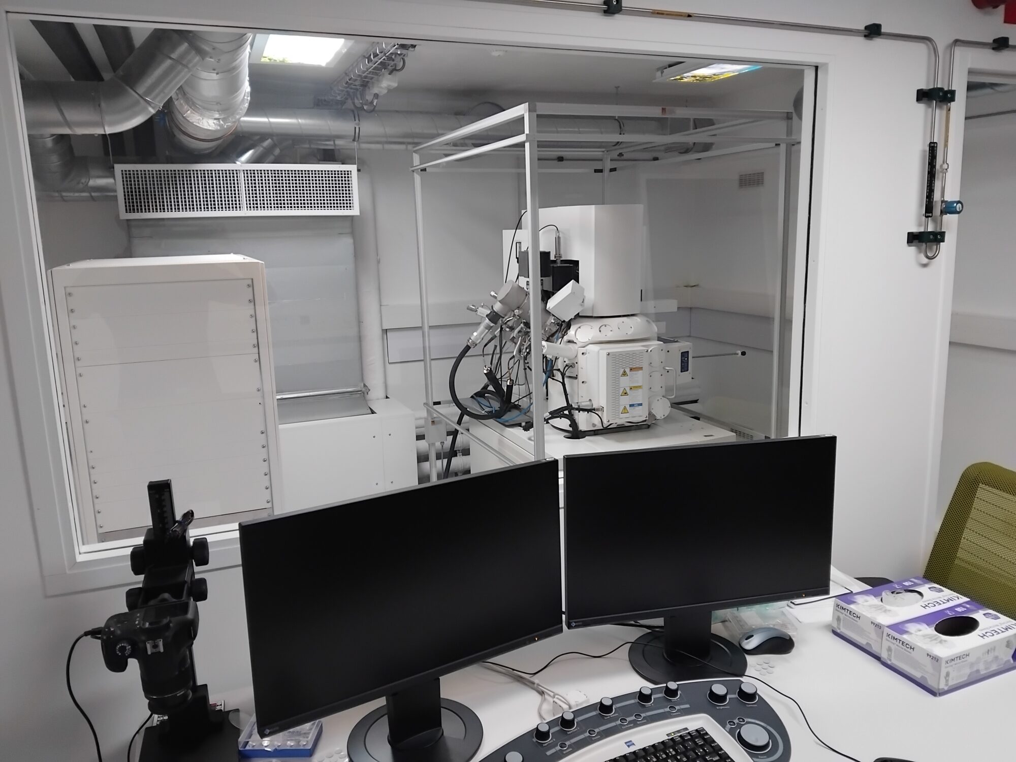
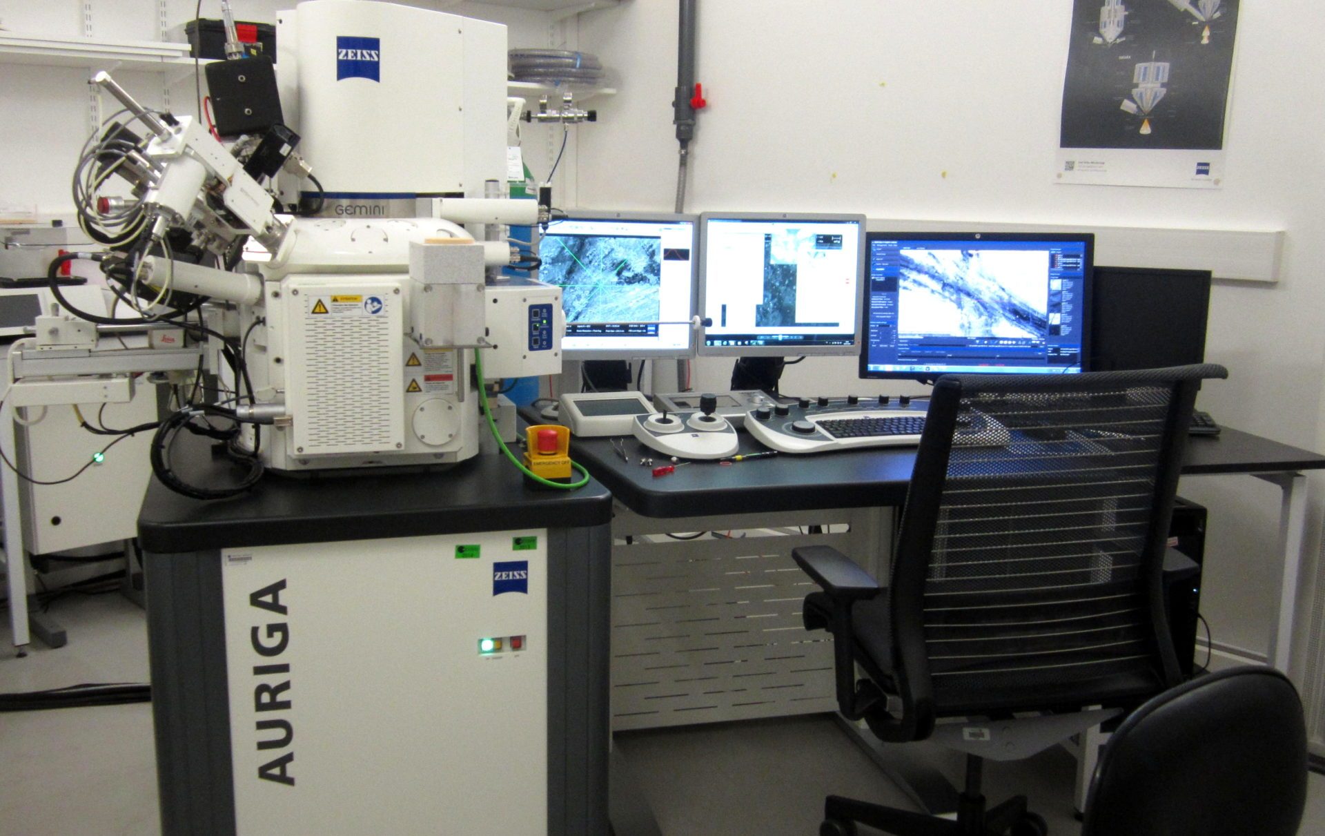
FIB-SEM cross beam, latest generation Carl Zeiss installed in 2025. FIB-SEM (Focused Ion Beam – Scanning Electron Microscopy) is a technique widely used in volumeEM (Electron Microscopy) to reconstruct high-resolution biological structures in 3D. The focused ion beam (gallium) is used to mill (remove very thin layers from the sample block, 3 to 10 nm thick). After each ablation, the SEM takes an image of the newly exposed surface. The sequence of images forms a stack of serial sections that can be reconstructed in 3D. FIB-SEM is now one of the benchmark techniques for EM volume imaging because it combines high-resolution isotropic imaging, automation, and accurate 3D reconstruction of cellular or tissue volumes.
This FIB-SEM is equipped with a Sculptor column, two backscattered electron detectors, and secondary electron detectors.