Search anything and hit enter
  • Teams
  • Members
  • Projects
  • Events
  • Calls
  • Jobs
  • publications
  • Software
  • Tools
  • Network
  • Equipment

A little guide for advanced search:

  • Tip 1. You can use quotes "" to search for an exact expression.
    Example: "cell division"
  • Tip 2. You can use + symbol to restrict results containing all words.
    Example: +cell +stem
  • Tip 3. You can use + and - symbols to force inclusion or exclusion of specific words.
    Example: +cell -stem
e.g. searching for members in projects tagged cancer
Search for
Count
IN
OUT
Content 1
  • member
  • team
  • department
  • center
  • program_project
  • nrc
  • whocc
  • project
  • software
  • tool
  • patent
  • Administrative Staff
  • Assistant Professor
  • Associate Professor
  • Clinical Research Assistant
  • Clinical Research Nurse
  • Clinician Researcher
  • Department Manager
  • Dual-education Student
  • Full Professor
  • Honorary Professor
  • Lab assistant
  • Master Student
  • Non-permanent Researcher
  • Nursing Staff
  • Permanent Researcher
  • Pharmacist
  • PhD Student
  • Physician
  • Post-doc
  • Prize
  • Project Manager
  • Research Associate
  • Research Engineer
  • Retired scientist
  • Technician
  • Undergraduate Student
  • Veterinary
  • Visiting Scientist
  • Deputy Director of Center
  • Deputy Director of Department
  • Deputy Director of National Reference Center
  • Deputy Head of Facility
  • Director of Center
  • Director of Department
  • Director of Institute
  • Director of National Reference Center
  • Group Leader
  • Head of Facility
  • Head of Operations
  • Head of Structure
  • Honorary President of the Departement
  • Labex Coordinator
Content 2
  • member
  • team
  • department
  • center
  • program_project
  • nrc
  • whocc
  • project
  • software
  • tool
  • patent
  • Administrative Staff
  • Assistant Professor
  • Associate Professor
  • Clinical Research Assistant
  • Clinical Research Nurse
  • Clinician Researcher
  • Department Manager
  • Dual-education Student
  • Full Professor
  • Honorary Professor
  • Lab assistant
  • Master Student
  • Non-permanent Researcher
  • Nursing Staff
  • Permanent Researcher
  • Pharmacist
  • PhD Student
  • Physician
  • Post-doc
  • Prize
  • Project Manager
  • Research Associate
  • Research Engineer
  • Retired scientist
  • Technician
  • Undergraduate Student
  • Veterinary
  • Visiting Scientist
  • Deputy Director of Center
  • Deputy Director of Department
  • Deputy Director of National Reference Center
  • Deputy Head of Facility
  • Director of Center
  • Director of Department
  • Director of Institute
  • Director of National Reference Center
  • Group Leader
  • Head of Facility
  • Head of Operations
  • Head of Structure
  • Honorary President of the Departement
  • Labex Coordinator
Search

← Go to Research

Go back
Scroll to top
Share
© Marie-Christine Prévost, Anne Derbise
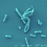
Bactéries Yersinia pestis en microscopie electronique à balayage.
Publication : PloS one

Imaging of bubonic plague dynamics by in vivo tracking of bioluminescent Yersinia pestis

Scientific Fields
Diseases
Organisms
Applications
Technique

Published in PloS one - 05 Apr 2012

Nham T, Filali S, Danne C, Derbise A, Carniel E

Link to Pubmed [PMID] – 22496846

PLoS ONE 2012;7(4):e34714

Yersinia pestis dissemination in a host is usually studied by enumerating bacteria in the tissues of animals sacrificed at different times. This laborious methodology gives only snapshots of the infection, as the infectious process is not synchronized. In this work we used in vivo bioluminescence imaging (BLI) to follow Y. pestis dissemination during bubonic plague. We first demonstrated that Y. pestis CO92 transformed with pGEN-luxCDABE stably emitted bioluminescence in vitro and in vivo, while retaining full virulence. The light produced from live animals allowed to delineate the infected organs and correlated with bacterial loads, thus validating the BLI tool. We then showed that the first step of the infectious process is a bacterial multiplication at the injection site (linea alba), followed by a colonization of the draining inguinal lymph node(s), and subsequently of the ipsilateral axillary lymph node through a direct connection between the two nodes. A mild bacteremia and an effective filtering of the blood stream by the liver and spleen probably accounted for the early bacterial blood clearance and the simultaneous development of bacterial foci within these organs. The saturation of the filtering capacity of the spleen and liver subsequently led to terminal septicemia. Our results also indicate that secondary lymphoid tissues are the main targets of Y. pestis multiplication and that colonization of other organs occurs essentially at the terminal phase of the disease. Finally, our analysis reveals that the high variability in the kinetics of infection is attributable to the time the bacteria remain confined at the injection site. However, once Y. pestis has reached the draining lymph nodes, the disease progresses extremely rapidly, leading to the invasion of the entire body within two days and to death of the animals. This highlights the extraordinary capacity of Y. pestis to annihilate the host innate immune response.