Link to Pubmed [PMID] – 12011015
Infect. Immun. 2002 Jun;70(6):3199-207
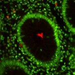
Shigella dysenteriae type 1-induced apoptotic cell death in rectal tissues from patients infected with Shigella dysenteriae type 1 was studied by the terminal deoxynucleotidyltransferase-mediated dUTP-biotin nick end labeling (TUNEL) technique and annexin V staining. Expression of proteins and cytokines participating in the apoptotic process (caspase-1, caspase-3, Fas [CD95], Fas ligand [Fas-L], perforin, granzyme A, Bax, WAF-1, Bcl-2, interleukin-2 [IL-2], IL-18, and granulocyte-macrophage colony-stimulating factor) in tissue in the acute and convalescent stages of dysentery was quantified at the single-cell level by in situ immunostaining. Apoptotic cell death in the lamina propria was markedly up-regulated at the acute stage (P < 0.05), where an increased number of necrotic cells were also seen. Phenotypic analysis of apoptotic cells revealed that 43% of T cells (CD3), 10% of granulocytes (CD15), and 5% of macrophages (CD56) underwent apoptosis. Increased activity of caspase-1 persisted in the rectum up to 1 month after onset. More-extensive expression of Fas, Fas-L, perforin, caspase-3, and IL-18, but not IL-2, at the acute stage than at the convalescent stage was observed. Increased expression of caspase-3 and IL-18 in tissues with severe inflammation compared to expression in those with mild inflammation was evident, implying a possible role in the perpetuation of inflammation. Significantly reduced cell death during convalescence was associated with a significant up-regulation of Bcl-2, Bax, and WAF-1 expression in the rectum compared to that in the acute phase of infection. Thus, induction of apoptosis at the local site in the early phase of S. dysenteriae type 1 infection was associated with a significant up-regulation of Fas/Fas-L and perforin and granzyme A expression and a down-regulation of Bcl-2 and IL-2, which promote cell survival.

